. 2020 Jun 26:11:388.
doi: 10.3389/fphys.2020.00388. eCollection 2020.

The Contribution of Surface Tension-Dependent Alveolar Septal Stress Concentrations to Ventilation-Induced Lung Injury in the Acute Respiratory Distress Syndrome

Affiliations

The Contribution of Surface Tension-Dependent Alveolar Septal Stress Concentrations to Ventilation-Induced Lung Injury in the Acute Respiratory Distress Syndrome

Carrie E Perlman. Front Physiol. .

Abstract

In the acute respiratory distress syndrome (ARDS), surface tension, T, is likely elevated. And mechanical ventilation of ARDS patients causes ventilation-induced lung injury (VILI), which is believed to be proportional to T. However, the mechanisms through which elevated T may contribute to VILI have been under-studied. This conceptual analysis considers experimental and theoretical evidence for static and dynamic mechanical mechanisms, at the alveolar scale, through which elevated T exacerbates VILI; potential causes of elevated T in ARDS; and T-dependent means of reducing VILI. In the last section, possible means of reducing T and improving the efficacy of recruitment maneuvers during mechanical ventilation of ARDS patients are discussed.

Keywords: acute respiratory distress syndrome (ARDS); mechanical ventilation; recruitment maneuvers; stress concentrations; surface tension; ventilation-induced lung injury (VILI).


Figures

FIGURE 1
FIGURE 1
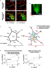
Single alveolar edema model. (A) Inflation and flooding effects on alveolar geometry. Optical sections of two surface alveoli in isolated, perfused rat lungs. Epithelium labeled with calcein red-orange. Transpulmonary pressure, PL, as indicated. In bottom images, right alveolus flooded with fluorescein-labeled 5% albumin solution. Air appears black. Sections are 2-μm thick and at 20-μm sub-pleural depth. Flooding right alveolus causes bowing of central “intervening” septum that separates aerated and flooded alveoli (arrows). Scale bars from baseline image are superimposed on all other images. Right alveolus that becomes flooded and left alveolus that remains aerated have areas AEdem and AAdj, respectively. (B) Meniscus in flooded alveolus. Image is y–z section, constructed from confocal z-stack, of an alveolus flooded with fluorescein-labeled 5% albumin solution. Pleural surface is at top of image. Epithelium is unlabeled and, along with air, appears black. Dashed line shows meniscus at mouth of flooded alveolus. (C) Schematics of alveolus before (left) and after (right) liquid flooding. Intervening septa separate aerated from flooded alveoli. Radiating septa are directed outward from flooded alveolus and have aerated alveoli to each side. PALV is alveolar air pressure. PLIQ is flooding liquid pressure. Air–liquid interface in flooded alveolus forms a meniscus. ΔPM = PALVPLIQ is pressure drop across meniscus. ΔPS = PALVPLIQ is pressure drop across intervening septa. Axial distending force FA acts at ends of sectioned septa and is greater in radiating septa around flooded alveolus on the right than in normally-stressed septa around aerated alveolus on the left. Ratio of summed FA over alveolar surface is an effective distending pressure PD applied to central alveolus. (D) Alveolar compliance. Alveolar areas plotted vs. PL for pairs of adjacent alveoli, one of which becomes flooded as in (A). Slope of lines is a two-dimensional analog of compliance. Statistics: Area greater at PL of 15 than 5 cm H2O (p < 0.01), for state before or after one alveolus is flooded. #Area different after than before one alveolus is flooded, at constant PL (p < 0.01). ##Same as “#” but p < 0.02. Slope less after than before one alveolus is flooded (p < 0.01). (A,B,D) Modified, with permission, from Perlman et al. (2011).
FIGURE 2
FIGURE 2
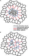
Stress concentrations in injured regions. Schematic representations showing stressed intervening and radiating septa in (A) homogeneously and (B) heterogeneously flooded regions. While only single layers of radiating septa are shown, computational modeling indicates radiating septa, especially those around a larger injured area, penetrate further into surrounding, aerated parenchyma (see text).
FIGURE 3
FIGURE 3
Forces acting on septa. (A) Normal planar septum between two aerated alveoli. Distending force FA applies axial tension. Interfacial surface tension, T, and tissue stress, σT, counter FA. (B) Intervening septum separating flooded (top) from aerated (bottom) alveolus. Due to ΔPS, which is ∼T, septum bows into flooded alveolus. Inflation-induced increases in FA and σT tend to reduce bowing. Inflation-induced increase in T acting along septum tends to reduce bowing, but inflation-induced increase in T also increases ΔPS, which tends to increase bowing. Net result is that inflation sometimes increases bowing (Figure 1A).
FIGURE 4
FIGURE 4
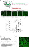
Ventilation-induced lung injury (VILI) assays. (A) Local VILI assay of ventilation-induced injury to alveolar–capillary barrier in the presence of heterogeneous alveolar flooding. Top schematic: Isolated lung is perfused with fluorescein-labeled blood/5% albumin mixture. A glass micropipette is used to puncture a surface alveolus and infuse non-fluorescent 3% albumin solution, which causes heterogeneous alveolar flooding in a local region. Menisci are present in flooded alveoli. Middle images: With PL held at 5 cm H2O, the region is imaged by confocal microscopy over 5-min baseline period. “L” labels alveoli flooded with non-fluorescent liquid. Then, five ventilation cycles are administered with an end-expiratory PL equivalent to an in vivo positive end-expiratory pressure (PEEP) of 10 cm H2O and a tidal volume, VT, of 12 ml/kg. Following ventilation, PL is returned to 5 cm H2O, and the region is imaged for 10 additional minutes. Bottom graph: plot of increase above baseline of normalized fluorescence in alveolar liquid vs. time. Gray: experimental, heterogeneously flooded regions in lungs subjected to five ventilation breaths with 10 cm H2O PEEP and VT as specified (fluorescein intensity quantified in alveolar flooding liquid). Black: control, aerated regions in lungs subjected to five ventilation breaths with 10 cm H2O PEEP and 6 or 12 ml/kg VT (data for two VT groups combined; fluorescein intensity quantified in liquid lining layer). Statistics: p < 0.01 vs. baseline; #p < 0.01 vs. both other groups at the same time point; °p < 0.01 vs. control group at the same time point. Figure modified, with permission, from Wu et al. (2014). (B) Global VILI assay of ventilation-induced increase in T in the presence of heterogeneous alveolar flooding. Table: summary of experimental protocol and T-values, all determined at PL of 15 cm H2O, at different time points. Statistics: p < 0.05 vs. T-values at two earlier time points. Image: representative view of alveolar flooding pattern on surface of lungs at final time point after induction of VILI and elevation of T. Alveolar edema liquid labeled by fluorescein administered to the perfusate. Figure modified, with permission, from Wu et al. (2017).
FIGURE 5
FIGURE 5
Pressure barrier, ΔPB, traps liquid in flooded alveoli. According to the Laplace relation, pressure in liquid below meniscus is PLIQ = PALV – 2T/R < PALV, where R is the meniscus radius. At the border between two adjacent alveoli, shared septum terminates at alveolar duct or sac, and liquid phases of alveoli are presumably continuous. Thus, in the plane of the figure, the interface has a small convex radius RB at the border of the flooded alveolus. In the plane perpendicular to the figure, the end of the septum and the interface should have larger concave radius RD of duct or sac, indicated by dashed gray curve. Again, according to the Laplace relation, the liquid phase pressure at the border of the alveolus is PLIQ.BORD = PALV + T(1/RB – 1/RD) > PALV. Thus, pressure barrier ΔPB = PLIQ.BORDPLIQ = T(1/RB – 1/RD + 2/R) > 0 traps liquid in the flooded alveolus. As ΔPBT, lowering T lowers ΔPB and increases the likelihood of liquid escape from the flooded alveolus, i.e., the likelihood of re-aeration. Figure modified, with permission, from Kharge et al. (2015) and Wu et al. (2017).

References

    1. Albert R. K., Smith B., Perlman C. E., Schwartz D. A. (2019). Is progression of pulmonary fibrosis due to ventilation-induced lung injury? Am. J. Respir. Crit. Care Med. 200 140–151. 10.1164/rccm.201903-0497PP - DOI - PMC - PubMed
    1. Anzueto A., Baughman R. P., Guntupalli K. K., Weg J. G., Wiedemann H. P., Raventós A. A., et al. (1996). Aerosolized surfactant in adults with sepsis-induced acute respiratory distress syndrome. Exosurf acute respiratory distress syndrome sepsis study group. N. Engl. J. Med. 334 1417–1421. 10.1056/NEJM199605303342201 - DOI - PubMed
    1. Arnal J.-M., Paquet J., Wysocki M., Demory D., Donati S., Granier I., et al. (2011). Optimal duration of a sustained inflation recruitment maneuver in ARDS patients. Intensive Care Med. 37 1588–1594. 10.1007/s00134-011-2323-0 - DOI - PubMed
    1. Bachofen H., Schürch S., Michel R. P., Weibel E. R. (1993). Experimental hydrostatic pulmonary edema in rabbit lungs. Morphology. Am. Rev. Respir. Dis. 147 989–996. 10.1164/ajrccm/147.4.989 - DOI - PubMed
    1. Bachofen H., Schürch S., Urbinelli M., Weibel E. R. (1987). Relations among alveolar surface tension, surface area, volume, and recoil pressure. J. Appl. Physiol. 62 1878–1887. 10.1152/jappl.1987.62.5.1878 - DOI - PubMed

LinkOut - more resources