. 2020 Jun 3:11:1157.
doi: 10.3389/fimmu.2020.01157. eCollection 2020.

The Receptor for Advanced Glycation Endproducts (RAGE) Contributes to Severe Inflammatory Liver Injury in Mice

Affiliations

The Receptor for Advanced Glycation Endproducts (RAGE) Contributes to Severe Inflammatory Liver Injury in Mice

Toni Weinhage et al. Front Immunol. .

Abstract

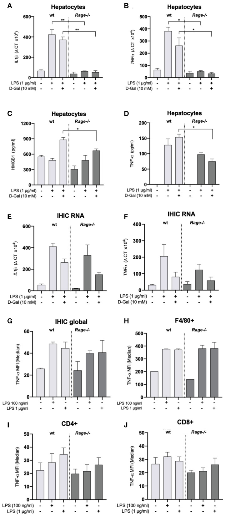
Background: The receptor for advanced glycation end products (RAGE) is a multiligand receptor involved in a number of processes and disorders. While it is known that RAGE-signaling can contribute to toxic liver damage and fibrosis, its role in acute inflammatory liver injury and septic multiorgan failure is yet undefined. We examined RAGE in lipopolysaccharide (LPS) induced acute liver injury of D-galN sensitized mice as a classical model for tumor necrosis factor alpha (TNF-α) dependent inflammatory organ damage. Methods: Mice (Rage-/- and C57BL/6) were intraperitoneally injected with D-galN (300 mg/kg) and LPS (10 μg/kg). Animals were monitored clinically, and cytokines, damage associated molecular pattern molecules (DAMPs) as well as liver enzymes were determined in serum. Liver histology, hepatic cytokines as well as RAGE mRNA expression were analyzed. Cellular activation and functionality were evaluated by flow cytometry both in bone marrow- and liver-derived cells. Results: Genetic deficiency of RAGE significantly reduced the mortality of mice exposed to LPS/D-galN. Hepatocyte damage markers were reduced in Rage-/- mice, and liver histopathology was less severe. Rage-/- mice produced less pro-inflammatory cytokines and DAMPs in serum and liver. While immune cell functions appeared normal, TNF-α production by hepatocytes was reduced in Rage-/- mice. Conclusions: We found that RAGE deletion attenuated the expression of pro-inflammatory cytokines and DAMPs in hepatocytes without affecting cellular immune functions in the LPS/D-galN model of murine liver injury. Our data highlight the importance of tissue-specific RAGE-signaling also in acute inflammatory liver stress contributing to sepsis and multiorgan failure.

Keywords: DAMPs; PAMPs; RAGE; inflammatory liver injury; sepsis.


Figures

Figure 1
Figure 1
Outcome of Rage–/– and wildtype (wt) mice after LPS/D-galN challenge. Mice were injected i.p. with 300 mg/kg D-galN and 10 μg/kg LPS. (A) Mouse survival post-injection is shown (n = 20, respectively, for each genotype in 3 independent experiments). (B) Serum levels of proinflammatory cytokines TNF-α, S100A9 as well as liver enzymes ALT and AST were measured immediately after mice succumbed to LPS/D-galN induced inflammatory liver injury (n = 3–4). (C) Wildtype (wt, light gray circles) and Rage–/– mice (dark gray circles, n = 4 each time point) were sacrificed after LPS/D-galN challenge at the time points indicated. Serum levels of TNF-α, IL-1β, MCP1, Interleukin-6, HMGB-1, IFN-γ, IL-1α, IL-12p70, IL-10, GM-CSF, IFN-β, IL-27, and IL-17A. The latter was not detectable. Data are expressed as the mean (± SEM). Statistical analysis was performed using Log rank (Mantel-Cox) (A) or Mann-Whitney test (B,C) comparing wt to Rage–/– mice. *p < 0.05, **p < 0.01, ***p < 0.001.
Figure 2
Figure 2
Hepatic pathology after LPS/D-galN challenge. (A) Histopathology in LPS-induced liver damage of D-galN sensitized Rage–/– and wildtype (wt) mice. H&E staining of wt and Rage–/– livers at 50x (upper panel, scale bar = 500 μm) and 400x (lower panel, scale bar = 50 μm) original magnifications. Massive necrosis, associated with intralobular hemorrhage (arrows), destruction of hepatic architecture as well as hepatocellular apoptosis (open arrows) and infiltration of neutrophils (red arrowheads) was visible in WT animals. Slight hepatic necrosis with minor inflammatory cell infiltration was observed in Rage–/– mice. (B) Quantification of LPS/D-galN induced inflammatory liver injury using a damage score that combines grading of hepatocellular necrosis, small vacuolisation and/or cell lysis, accumulation of erythrocytes in the sinusoids and neutrophilic infiltration at the time points indicated (n = 4 each time point). (C) Rage mRNA expression in wt liver tissue at 2 and 4 h after LPS/D-galN injection (n = 3). (D) Liver mRNA expression of pro-inflammatory cytokines Tnf-α, Interleukin-1 beta, and Interleukin-6 in mice after 4 h of LPS/D-galN induced inflammatory liver injury (n = 4–5). Data represent the mean + SEM of the results obtained from two independent experiments. Statistical analysis was performed using Mann-Whitney test comparing wildtype to Rage–/– mice (B,D) or untreated to challenged wildtype mice (C), *p < 0.05, **p < 0.01. LPS, lipopolysaccharide; D-Gal, D-galN (D-galactosamine).
Figure 3
Figure 3
Localization of DAMPs in hepatic tissue. (A) Analyses in non-challenged wildtype (wt) or Rage–/– mice. Immunohistochemical staining at 200x (upper panels, scale bars 50 μM) confirmed the expression of HMGB-1 in parenchymal and non-parenchymal hepatic cells, which was restricted to the nucleus. S100A8 was only found in the cytoplasm of few Kupffer cells (white open arrows). Immunofluorescence analyses at 400x (lower panel, scale bars 50 μM) confirmed the distinct staining patterns of HMGB-1 (FITC, green) and S100A8 (Alexa fluor 488, red). DAPI (blue) was used to counterstain nuclei. (B) In the liver of mice 4 h after LPS/D-galN injection, there was a stronger staining of HMGB-1, but without significant translocation from the nucleus into the cytoplasm within the short timeframe. In addition, more infiltrating S100A8-positive myeloid cells were present (white open arrows). When compared to Rage–/– animals, the expression of DAMPs was more pronounced in damaged liver sections from wt mice.
Figure 4
Figure 4
Functional characterization of neutrophils and bone marrow derived monocytes/macrophages (BMDM). (A) ROS production of neutrophils from wildtype (wt) or Rage–/– mice after 1 h. (B) Phagocytosis of FITC-labeled E. coli by neutrophils after 1 h. (C) NETosis of neutrophils after treatment with ionomycin (4 μM) for 60–180 min. (D) ROS production of BMDM after 1 h. (E) Phagocytosis of FITC-labeled E. coli by BMDM after 1 h. (F) Release TNF-α by BMDM into the cell culture supernatant after 6 h without (w/o) or with LPS (100 ng) stimulation. Data are expressed as the mean + SEM. MFI, mean fluorescence intensity; ROS, reactive oxygen species.
Figure 5
Figure 5
Characterization of hepatocytes and intrahepatic immune cells (IHICs). Primary liver cells were generated by established protocols. Cells were further isolated to separate hepatocytes and IHICs. (A–D) After culture for 4 days, hepatocytes were treated for 4 h with LPS (1 μg/ml) and D-galN (10 mM) or left untreated. Relative mRNA expression of Il-1β (A) and Tnf(B) in hepatocytes quantified by qRT-PCR. Gene expression levels were normalized to two housekeeper genes (Hprt and Rps9). (C) HMGB1 and (D) TNF-α concentrations released from hepatocytes into the supernatants after in vitro stimulation of hepatocytes with LPS and D-galN for 4 h. The stimulation with D-galN alone had no effect (not shown). Data are expressed as the mean ± SEM of two experiments (n = 4 per group). IHICs were isolated and subsequently stimulated with LPS for 4 h. Relative mRNA expression of Il-1β (E) and Tnf(F) in hepatocytes was quantified by qRT-PCR. Gene expression levels were normalized to two housekeeper genes (Hprt and Rps9). (G,H) IHICs were isolated and subsequently stimulated with LPS for 4 h in the presence of a protein transport inhibitor, followed by intracellular staining and FACS analyses to quantify intracellular TNF-α production. (G) Quantification of intracellular TNF-α production in IHIC isolated from Rage–/– and wildtype (wt) mice. (H) Quantification of intracellular TNF-α production in F4/80+/CD11b+ cells (resident macrophages/Kupffer cells). (I,J) Quantification of intracellular TNF-α production in CD4+ or CD8+ T lymphocytes. Data are depicted as mean ±SEM of two experiments (n = 4 per group). Data are expressed as the median ± SEM of two experiments (n = 4 per group). Statistical analysis was performed using one-way ANOVA; *p ≤ 0.05, **p ≤ 0.01. LPS, lipopolysaccharide; D-Gal, D-galN, D-galactosamine.

References

    1. Engel C, Brunkhorst FM, Bone HG, Brunkhorst R, Gerlach H, Grond S, et al. . Epidemiology of sepsis in Germany: results from a national prospective multicenter study. Intensive Care Med. (2007) 33:606–18. 10.1007/s00134-006-0517-7 - DOI - PubMed
    1. van der Poll T, van de Veerdonk FL, Scicluna BP, Netea MG. The immunopathology of sepsis and potential therapeutic targets. Nat Rev Immunol. (2017) 17:407–20. 10.1038/nri.2017.36 - DOI - PubMed
    1. Rubio I, Osuchowski MF, Shankar-Hari M, Skirecki T, Winkler MS, Lachmann G, et al. . Current gaps in sepsis immunology: new opportunities for translational research. Lancet Infect Dis. (2019) 19:e422–36. 10.1016/S1473-3099(19)30567-5 - DOI - PubMed
    1. Galanos C, Freudenberg MA, Reutter W. Galactosamine-induced sensitization to the lethal effects of endotoxin. Proc Natl Acad Sci USA. (1979) 76:5939–43. 10.1073/pnas.76.11.5939 - DOI - PMC - PubMed
    1. Silverstein R. D-galactosamine lethality model: scope and limitations. J Endotoxin Res. (2004) 10:147–62. 10.1179/096805104225004879 - DOI - PubMed

Publication types

Substances