Review
. 2020 Jun 24:11:1169.
doi: 10.3389/fimmu.2020.01169. eCollection 2020.

Crosstalk Between Liver Macrophages and Surrounding Cells in Nonalcoholic Steatohepatitis

Affiliations
Review

Crosstalk Between Liver Macrophages and Surrounding Cells in Nonalcoholic Steatohepatitis

Haiou Li et al. Front Immunol. .

Abstract

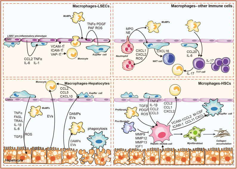
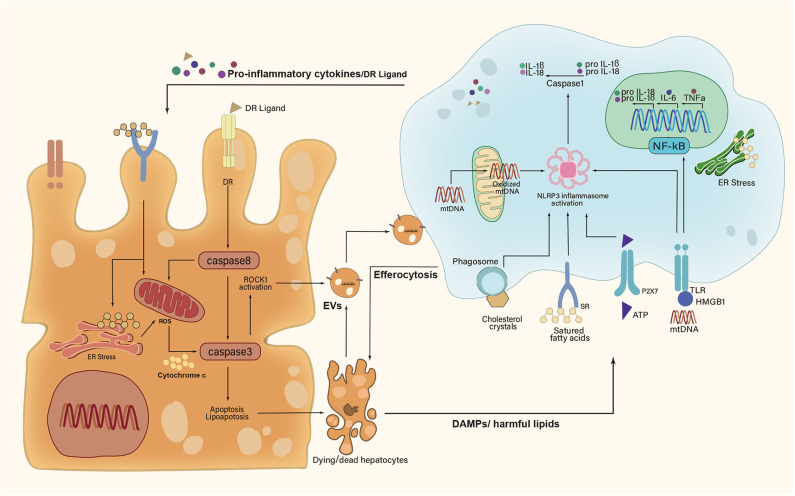
Nonalcoholic steatohepatitis (NASH), the advanced stage of nonalcoholic fatty liver disease (NAFLD), is emerging as a leading cause of progressive liver fibrosis and end-stage liver disease. Liver macrophages, mainly composed of Kupffer cells (KCs) and monocyte-derived macrophages (MoMFs), play a vital role in NASH progression and regression. Recent advances suggest that cell-cell communication is a fundamental feature of hepatic microenvironment. The reprogramming of cell-cell signaling between macrophages and surrounding cells contributes to the pathogenesis of NASH. In this review, we summarize the current knowledge of NASH regarding the composition of liver macrophages and their communication with surrounding cells, which are composed of hepatocytes, hepatic stellate cells (HSCs), liver sinusoidal endothelial cells (LSECs) and other immune cells. We also discuss the potential therapeutic strategies based on the level of macrophages.

Keywords: cellular crosstalk; liver cells; liver macrophages; nonalcoholic steatohepatitis; therapeutic strategies.


Figures

Figure 1
Figure 1
Overview of liver macrophage-related intercellular signaling in nonalcoholic steatohepatitis (NASH). The illustration consists of four groups, as follows: liver macrophages–hepatocytes; liver macrophages–hepatic stellate cells (HSCs); liver macrophages–liver sinusoidal endothelial cells (LSECs); liver macrophages–immune cells. DAMPs, damage-associated molecular patterns; EVs, extracellular vesicles; TNFα, tumor necrosis factor α; TRAIL, TNF-related apoptosis-inducing ligand; FasL, Fas ligand; ROS, reactive oxygen species; CCL, chemokine (C-C) motif ligand; CXCL, chemokine (C-X-C motif) ligand; IL, interleukin; MMP, matrix metalloproteinase; IGF1, insulin-like growth factor 1; TGFβ, transforming growth factor-β; M-CSF, macrophage colony-stimulating factor; PDGF, platelet-derived growth factor; PAF, platelet-activating factor; ICAM-1, intercellular adhesion molecule-1; VCAM-1, vascular cell adhesion molecule-1; VAP-1, vascular adhesion protein-1; MPO, myeloperoxidase; NO, nitric oxide; IFNγ, interferon γ.
Figure 2
Figure 2
A feed-forward regulatory loop between lipotoxic hepatocytes and Kupffer cells. Upon metabolic stress, dying and dead hepatocytes release damage-associated molecular patterns (DAMPs), extracellular vesicles (EVs), and harmful lipids to activate Kupffer cells (KCs). In turn, activated KCs secrete proinflammatory cytokines and death receptor (DR) ligands to aggravate hepatocyte damage. However, KCs can remove apoptotic hepatocytes via efferocytosis. IL-1β, interleukin-1β; IL-6, interleukin-6; IL-18, interleukin-18; TNFα, tumor necrosis factor α; HMGB1, high mobility group box-1; ATP, adenosine triphosphate; mtDNA, mitochondrial DNA; ROCK1, rho-associated, coiled-coil containing protein kinase 1; NF-κB, nuclear factor-κB; NLRP3, NLR family pyrin domain-containing 3; ER, endoplasmic reticulum; ROS, reactive oxygen species; P2X7, P2X purinoceptor 7; TLR, toll-like receptor; SR, scavenger receptor.

References

    1. Di Sessa A, Cirillo G, Guarino S, Marzuillo P, Miraglia Del Giudice E. Pediatric non-alcoholic fatty liver disease: current perspectives on diagnosis and management. Pediatric Health Med Ther. (2019) 10:89–97. 10.2147/PHMT.S188989 - DOI - PMC - PubMed
    1. Diehl AM, Day C. Cause, pathogenesis, and treatment of nonalcoholic steatohepatitis. N Engl J Med. (2017) 377:2063–72. 10.1056/NEJMra1503519 - DOI - PubMed
    1. Doycheva I, Issa D, Watt KD, Lopez R, Rifai G, Alkhouri N. Nonalcoholic steatohepatitis is the most rapidly increasing indication for liver transplantation in young adults in the united states. J Clin Gastroenterol. (2018) 52:339–46. 10.1097/MCG.0000000000000925 - DOI - PubMed
    1. Sheka AC, Adeyi O, Thompson J, Hameed B, Crawford PA, Ikramuddin S. Nonalcoholic steatohepatitis: a Review. JAMA. (2020) 323:1175–83. 10.1001/jama.2020.2298 - DOI - PubMed
    1. Kazankov K, Jorgensen SMD, Thomsen KL, Moller HJ, Vilstrup H, George J, et al. . The role of macrophages in nonalcoholic fatty liver disease and nonalcoholic steatohepatitis. Nat Rev Gastroenterol Hepatol. (2019) 16:145–59. 10.1038/s41575-018-0082-x - DOI - PubMed

Publication types

MeSH terms