. 2020 May 27:11:645.
doi: 10.3389/fpls.2020.00645. eCollection 2020.

Development of a Direct in vitro Plant Regeneration Protocol From Cannabis sativa L. Seedling Explants: Developmental Morphology of Shoot Regeneration and Ploidy Level of Regenerated Plants

Affiliations

Development of a Direct in vitro Plant Regeneration Protocol From Cannabis sativa L. Seedling Explants: Developmental Morphology of Shoot Regeneration and Ploidy Level of Regenerated Plants

Alberto Galán-Ávila et al. Front Plant Sci. .

Abstract

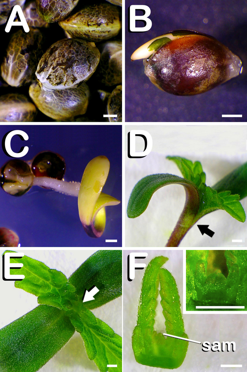
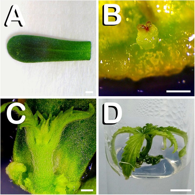
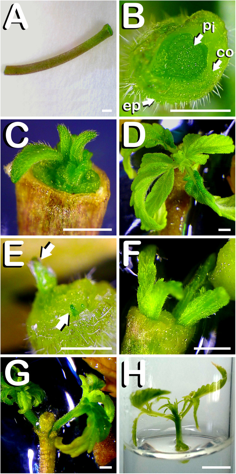
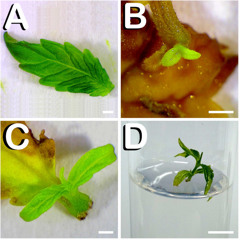
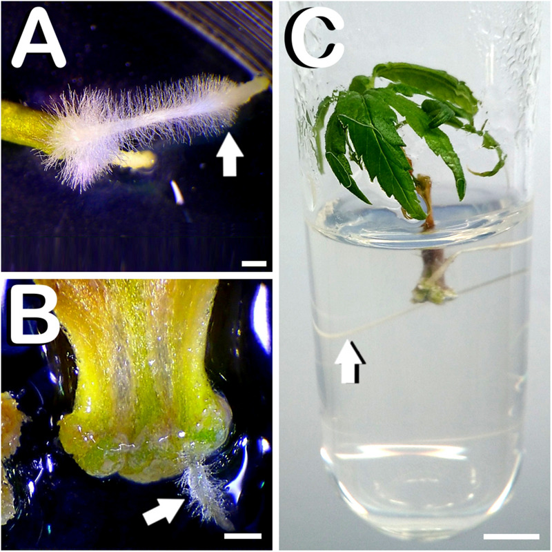
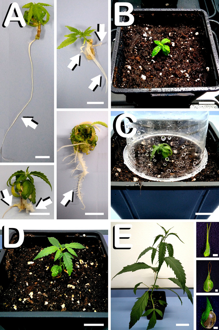
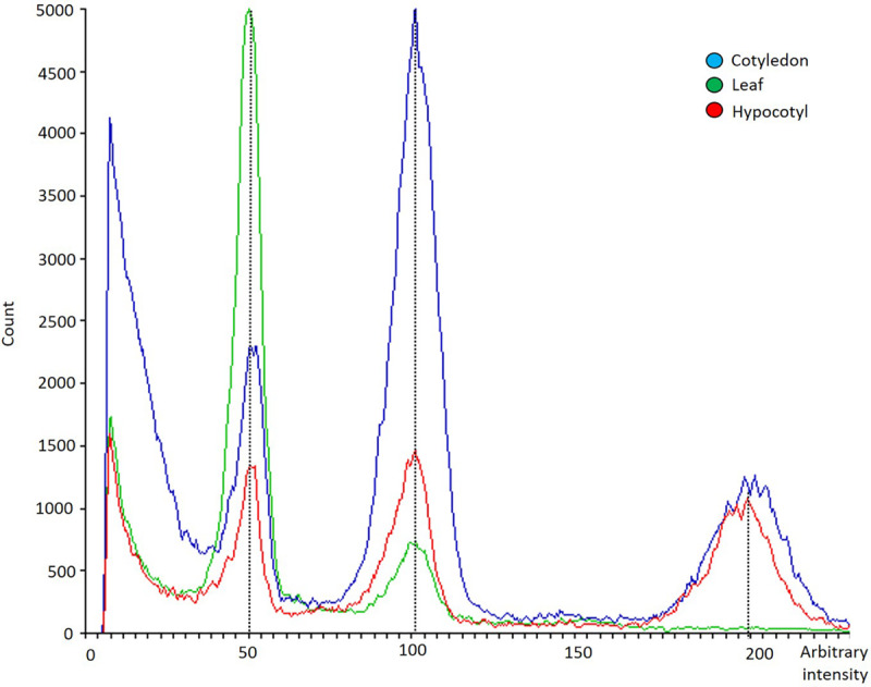
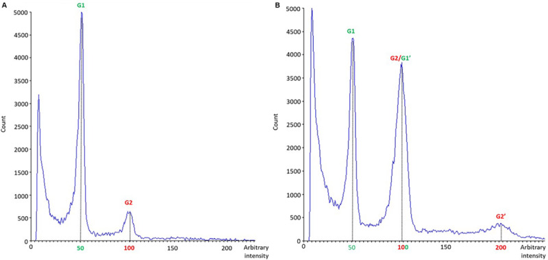
In vitro shoot regeneration can efficiently contribute to the improvement of recalcitrant Cannabis sativa L. We aimed at developing a highly efficient protocol for in vitro direct regeneration of C. sativa plants from different explants (cotyledon, hypocotyl, and true leaf) from seedlings of monoecious C. sativa short-day varieties Ferimon, Felina32, Fedora17, and USO31, together with dioecious neutral-day variety Finola. Ten regeneration media, including already published protocols, and self-designed combinations of plant growth regulators were tested. The developmental morphology since germination of seeds to the development of rooted plantlets was followed. Additionally, the ploidy level of explants and in vitro regenerants was analyzed. We concluded that hypocotyl is the best explant for in vitro direct regeneration of C. sativa plants with 49.45% of responding explants, while cotyledon and true leaf had a poor response with, respectively, 4.70 and 0.42% of explants developing plantlets. In terms of shoot regeneration, we found significant differences among the culture media evaluated and the varieties studied. Overall, the best regeneration media were ZEARIB 2.0 (mg/L) and ZEARIB 1.0 (mg/L) + NAA 0.02 (mg/L) with 66.67% of responding hypocotyls. Amazingly, hypocotyls cultured in medium without plant growth regulators showed an excellent response (61.54% of responding hypocotyls) and spontaneous rooting of regenerants (17.94%). In vitro regenerated plants were acclimatized just 6 weeks after culture initiation. The developmental morphology study suggests that regenerated shoots originate from pericycle cells adjacent to xylem poles. Polysomaty was detected in hypocotyls and cotyledons of all varieties studied, and diploid (>80%) and mixoploid (with diploid and tetraploid cells) plants were regenerated. Our protocol allows a high shoot organogenesis efficiency in different C. sativa varieties. The fact that a significant percentage of plants are mixoploid may provide an alternative way to develop polyploids in C. sativa. Our results show that direct in vitro regeneration may make a significant contribution to the development of improved C. sativa materials for medical applications.

Keywords: cannabinoids; hemp; hypocotyl; micropropagation; polyploidization; polysomaty; shoot organogenesis.


Figures

FIGURE 1
FIGURE 1
Seed germination of C. sativa. The different developmental stages are described as follows: (A) Seeds just before being sterilized. (B) Germinated seed 48 h. after in vitro sowing with the root apical meristem arising from testa. (C) Emerging seedling 5 days after seed plating, with testa being visible at the bottom of the image. (D) Seven-days-old seedling with fully expanded first pair of true leaves, which is equivalent to the phenological growth stage coded in this species by number 11 in BBCH-scale: arrow marks dissection point. (E) View of 7-days-old seedling allowing observation of vegetative shoot apex: arrow points shoot apex location on seedling. (F) Remaining vegetative shoot apex after dissection of hypocotyl, cotyledon and true leaves from 7 days-old seedling, with shoot apical meristem (SAM) highlighted on it: detail of SAM (inset in panel F). Scale bars: 1 mm.
FIGURE 2
FIGURE 2
Direct in vitro shoot organogenesis from cotyledon leaves of C. sativa. The different developmental stages are described as follows: (A) Newly dissected cotyledon leaf from a 7-days-old hemp seedling. (B) Shoot primordium formation at the basal zone of cotyledon leaf after 4 days of in vitro culture. (C) Vigorous shoot arising from the lower part of cotyledon leaf 14 days after exposure to the culture medium. (D) Cotyledon derived plant cultured in a glass-tube 21 days after explant inoculation. Scale bars (A–C): 1 mm. Scale bar (D): 6 mm.
FIGURE 3
FIGURE 3
Direct in vitro shoot organogenesis from hypocotyls of C. sativa. The different developmental stages are described as follows: (A) Newly dissected hypocotyl from a 7-days-old hemp seedling. (B) Transverse section of newly dissected hemp hypocotyl revealing its different layers: ep: epidermis; co: cortex; pi: pith. (C) Formation of one shoot at the top of the hypocotyl after 7 days of in vitro culture. (D) Vigorous shoot arising from the upper part of hypocotyl 14 days after exposure to the culture medium. (E) Two primordia arising from the top of the hypocotyl after 4 days of in vitro culture: arrows point both primordia. (F) Two hypocotyl derived plants 9 days after explant inoculation. (G) Two hypocotyl derived regenerants ready to be subcultured 14 days after explant culture. (H) Hypocotyl derived plant individually grown in a glass-tube 21 days after culture initiation. Scale bars (A–G): 1 mm. Scale bar (H): 6 mm.
FIGURE 4
FIGURE 4
Direct in vitro shoot organogenesis from true leaves of C. sativa. The different developmental stages are described as follows: (A) Newly dissected leaf from a 7-days-old hemp seedling. (B) Formation of one primordium from leaf-petiole transition zone 1 week after culture initiation. (C) Two-week-old plantlet of approximately one centimeter in height ready for subculture. (D) Leaf derived plant individually grown in a glass-tube 21 days after culture initiation. Scale bars (A–C): 1 mm. Scale bar (D): 6 mm.
FIGURE 5
FIGURE 5
Rooting of explants and spontaneous rooting of hypocotyl derived plants of C. sativa. (A) Vigorous root with radicular hairs emerging from the basal zone of the hypocotyl 2 weeks after culture initiation (arrow). (B) Small root with root hairs arising from the lower part of the cotyledon after 14 days of in vitro culture (arrow). (C) Spontaneously rooted hypocotyl derived plant after 28 days of culture initiation with a prominent root (arrow). Scale bars (A,B): 1 mm. Scale bar (C): 6 mm.
FIGURE 6
FIGURE 6
Acclimatization process of hypocotyl derived plants in C. sativa. The different developmental stages are described as follows: (A) Radicular system of hypocotyl derived plants spontaneously rooted 28 days after culture initiation, where different root morphogenesis patterns can be observed (arrows). (B) Small plant just after being transplanted to pots (2 L) with fertilized commercial substrate. (C) Plastic vessel covering the in vitro regenerated plant in order to avoid desiccation. (D) Hypocotyl derived plant exposed to the environmental humidity 6 weeks after culture initiation. (E) Female hypocotyl derived hemp plant showing sexual functionality 8 weeks after in vitro explant inoculation (insets illustrates, from top to bottom, unfertilized female flower, fertilized female flower during seed formation and mature seed final development). Scale bars (A–D): 12 mm. Scale bar (E): 60 mm. Scale bars of insets (E): 1 mm.
FIGURE 7
FIGURE 7
Flow cytometry histogram showing polysomatic pattern in cotyledons (blue), hypocotyls (red) and first pair of true leaves (green) from C. sativa. The x-axis represents a fluorescence intensity level proportional to the nuclear DNA content. The peak located at the value 50 corresponds to the diploid nuclei in phase G1, the peak located at the value 100 corresponds to the sum of the diploid nuclei in phase G2 and the tetraploid nuclei in phase G1, while the one at the value 200 represents tetraploid nuclei in G2 phase. The y-axis indicates the number of nuclei analyzed.
FIGURE 8
FIGURE 8
Nuclear DNA histogram patterns of diploid (A) and mixoploid (B) in vitro regenerated plants of C. sativa analyzed by flow cytometry. The x-axis represents a fluorescence intensity level proportional to the nuclear DNA content. The peak located at the value 50 corresponds to the diploid nuclei in phase G1, the peak located at the value 100 corresponds to the sum of the diploid nuclei in phase G2 and the tetraploid nuclei in phase G1, while the one at the value 200 represents tetraploid nuclei in G2 phase. The y-axis indicates the number of nuclei analyzed.

References

    1. Adelberg J. W., Rhodes, Bill B., Skorupska H. T. (1993). Generating tetraploid melons in tissue culture. Acta Hortic. 336 373–380. 10.17660/actahortic.1993.336.49 - DOI
    1. Andre C. M., Hausman J. F., Guerriero G. (2016). Cannabis sativa: the plant of the thousand and one molecules. Front. Plant Sci. 7:19 10.3389/fpls.2016.00019 - DOI - PMC - PubMed
    1. Atta R., Laurens L., Boucheron-Dubuisson E., Guivarc’h A., Carnero E., Giraudat-Pautot V., et al. (2009). Pluripotency of Arabidopsis xylem pericycle underlies shoot regeneration from root and hypocotyl explants grown in vitro. Plant J. 57 626–644. 10.1111/j.1365-313X.2008.03715.x - DOI - PubMed
    1. Beck E. H. (1996). Regulation of shoot/root ratio by cytokinins from roots in Urtica dioica: opinion. Plant Soil 185 3–12. 10.1007/bf02257560 - DOI
    1. Beeckman T., De Smet I. (2014). Pericycle. Curr. Biol. 24 R378–R379. 10.1016/j.cub.2014.03.031 - DOI - PubMed