. 2020 Jul 8:6:30.
doi: 10.1186/s40942-020-00231-2. eCollection 2020.

New findings useful for clinical practice using swept-source optical coherence tomography angiography in the follow-up of active ocular toxoplasmosis

Affiliations

New findings useful for clinical practice using swept-source optical coherence tomography angiography in the follow-up of active ocular toxoplasmosis

João Rafael de Oliveira Dias et al. Int J Retina Vitreous. .

Abstract

Background: Ocular toxoplasmosis is one of the most common causes of intraocular inflammation and posterior uveitis in immunocompetent patients. This paper aims to investigate swept-source optical coherence tomography angiography (SS-OCTA) findings in eyes with active toxoplasmic retinochoroiditis.

Methods: This case series was conducted from November 2017 through October 2019 in two Brazilian centers. 15 eyes of 15 patients with active toxoplasmic retinochoroiditis were included, and were imaged at baseline and after at least 4 weeks of follow-up. All patients underwent ophthalmic examinations and multimodal imaging including SS-OCT and SS-OCTA before and after treatment of ocular toxoplasmosis. The differential diagnoses included toxoplasmosis, syphilis, and human immunodeficiency virus, which were eliminated through serologic and clinical evaluations.

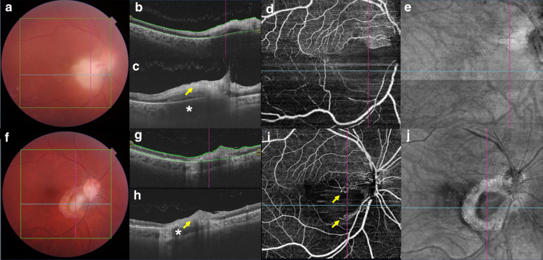
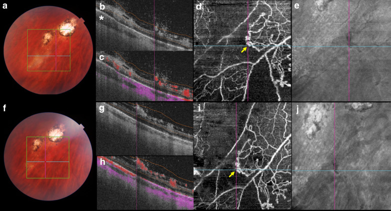
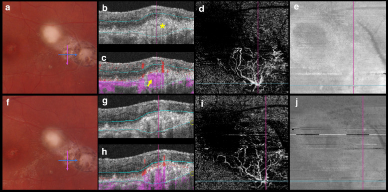
Results: All 15 patients presented with positive anti-Toxoplasma gondii immunoglobulin G titers and three also presented with positive anti-T. gondii immunoglobulin M titers. The mean age at examination was 32.4 years ± 12.7 years (range 15-59 years). Sixty percent of the patients were female. In all eyes, the inner retinal layers were abnormally hyperreflective with full-thickness disorganization of the retinal reflective layers at the site of the active toxoplasmic retinochoroiditis. At baseline, 80% of eyes had focal choroidal thickening beneath the retinitis area, and all eyes had a choroidal hyporeflective signal. Before treatment, SS-OCTA showed no OCTA decorrelation signal next to the lesion site in all eyes, and flow signal improvement was noticed after treatment. Three eyes presented with intraretinal vascular abnormalities during follow-up. SS-OCTA showed retinal neovascularization in one patient and a presumed subclinical choroidal neovascular membrane in another patient.

Conclusions: SS-OCT and SS-OCTA are useful for assessing unexpected structural and vascular retinal and choroidal changes in active and post-treatment toxoplasmic retinochoroiditis and these findings are useful for clinical practice.

Keywords: Ocular toxoplasmosis; Optical coherence tomography angiography; Toxoplasmic retinochoroiditis.


Conflict of interest statement

Competing interestsThe authors declare that they have no competing interests.

Figures

Fig. 1
Fig. 1
a Color fundus at the time of diagnosis. Vitritis and an active toxoplasmic lesion are visible in the papillomacular bundle. b Optical coherence tomography (OCT) B-scan with the total retina slabs. c OCT B-scan over the active lesion shows retinal hyperreflectivity (arrow) and a thickened choroid (asterisk) under the lesion. d A 9 × 9-mm field of view swept-source OCTA (SS-OCTA) image shows inferiorly located no OCTA decorrelation signal, suggestive of reduced blood flow. e Structural en face image. f Color fundus image 13 weeks after those in (ae): inflammatory signs are absent, but an atrophic scar is visible. g OCT B-scan with the total retina slabs. h Retinal layers are unrecognizable at the lesion site (arrow) and choroidal thinning is seen (asterisk). i A 9 × 9-mm field of view on SS-OCTA shows increase in flow signal after treatment. The arrows indicate the vascular loops. j A structural en face image
Fig. 2
Fig. 2
a Color fundus at the time of diagnosis. An active toxoplasmic lesion is visible adjacent to the retinal choroidal pigmented scars. b An optical coherence tomography (OCT) B-scan with slabs extending from the vitreous to the inner nuclear layer. Thickened choroidal tissue is seen adjacent to the lesion site (asterisk). c An OCT B-scan with overlying color-coded flow in which red represents the retinal capillaries and pre-retinal neovascularization, and pink represents the choroid. d A 6 × 6-mm field of view on OCT angiography (OCTA) shows a neovascular seafan-like complex (arrow) and areas of no decorrelation signal. e A structural en face OCT shows a hyporeflective lesion suggestive of retinal neovascularization. f A color fundus image obtained 5 weeks after those in (ae) shows that the area of retinitis has regressed following ocular toxoplasmosis treatment. g An OCT B-scan with slabs extending from the vitreous to the inner nuclear layer. h An OCT B-scan with overlying color-coded flow shows that the retinal neovascularization appears unchanged after the systemic anti-Toxoplasma treatment. i A 6 × 6-mm OCTA image shows unchanged retinal neovascularization (arrow). j A structural en face OCT image
Fig. 3
Fig. 3
a Color fundus 6 weeks after treatment. The pink and blue arrows indicate the B-scan positions in B and C. b An optical coherence tomography (OCT) B-scan with the outer retina slabs. Hyperreflective material is seen in the subretinal space (asterisk). c An OCT B-scan with overlying color-coded flow in which red represents the retinal capillaries, and pink represents the choroid and the choroidal neovascular membrane (CNVM). d A 3 × 3-mm field of view on a swept-source OCT angiography (SS-OCTA) image suggests the presence of a CNVM. e A structural en face OCT shows hyporeflective dots in the CNVM location. f Color fundus image 4 weeks after those in (ae). g An OCT B-scan with the outer retina slabs. h An OCT B-scan with overlying color-coded flow. i A 3 × 3-mm OCTA image showing the CNVM. j A structural en face image

References

    1. Furtado JM, Smith JR, Belfort R, Jr, Gattey D, Winthrop KL. Toxoplasmosis: a global threat. J Glob Infect Dis. 2011;3(3):281–284. - PMC - PubMed
    1. Nussenblatt RB, Belfort R., Jr Ocular toxoplasmosis. An old disease revisited. JAMA. 1994;271(4):304–307. - PubMed
    1. Jones JL, Dubey JP. Waterborne toxoplasmosis—recent developments. Exp Parasitol. 2010;124(1):10–25. - PubMed
    1. Montoya JG, Liesenfeld O. Toxoplasmosis. Lancet. 2004;363(9425):1965–1976. - PubMed
    1. Torgerson PR, Mastroiacovo P. The global burden of congenital toxoplasmosis: a systematic review. Bull World Health Organ. 2013;91(7):501–508. - PMC - PubMed

LinkOut - more resources