

The Future of Concurrent Automated Coronary Artery Calcium Scoring on Screening Low-Dose Computed Tomography
- PMID: 32670710
- PMCID: PMC7358941
- DOI: 10.7759/cureus.8574
The Future of Concurrent Automated Coronary Artery Calcium Scoring on Screening Low-Dose Computed Tomography
Abstract
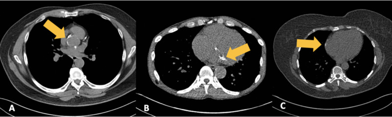
Low-dose computed tomography (LDCT) has been extensively validated for lung cancer screening in selected patient populations. Additionally, the use of gated cardiac CT to assess coronary artery calcium (CAC) burden has been validated to determine a patient's risk for major cardiovascular adverse events. This is typically performed by calculating an Agatston score based on density and overall burden of calcified plaque within the coronary arteries. Patients that qualify for LDCT for lung cancer screening commonly share major risk factors for coronary artery disease and would frequently benefit from an additional gated cardiac CT for the assessment of CAC. Given the widespread use of LDCT for lung cancer screening, we evaluated current literature regarding the use of non-gated chest CT, specifically LDCT, for the detection and grading of coronary artery calcifications. Additionally, given the evolving and increasing use of artificial intelligence (AI) in the interpretation of radiologic studies, current literature for the use of AI in CAC assessment was reviewed. We reviewed primary scientific literature dating up to April 2020 using Pubmed and Google Scholar, with the search terms low dose CT, lung cancer screening, coronary artery calcium, EKG/cardiac gated CT, deep learning, machine learning, and AI. These publications were then independently evaluated by each member of our team. Overall, there was a consensus within these papers that LDCT for lung cancer screening plays a role in the evaluation of CAC. Most studies note the inherent problems with the evaluation of the density of coronary calcifications on LDCT to give an accurate numeric calcium or Agatston score. The current method of evaluating CAC on LDCT involves using a qualitative categorical system (none, mild, moderate, or severe). When performed by cardiac imaging experts, this method broadly correlates with traditional CAC score groups (0, 1 to 100, 101 to 400, and > 400). Furthermore, given the high sensitivity of a properly protocolled LDCT for coronary calcium, a negative study for CAC precludes the need for a dedicated gated CT assessment. However, qualitative methods are not as accurate or reproducible when performed by general radiologists. The implementation of AI in the LDCT screening process has the potential to give a quantifiable and reproducible numeric value to the calcium score, based on whole heart volume scoring of calcium. This more closely aligns with the Agatston score and serves as a better guide for treatment and risk assessment using current guidelines. We conclude that CAC should be assessed on all LDCT performed for lung cancer screening and that a qualitative categorical scoring system should be provided in the impression for each patient. Early studies involving AI for the assessment of CAC are promising, but more extensive studies are needed before a final recommendation for its use can be given. The implementation of an accurate, automated AI CAC assessment tool would improve radiologist compliance and ease of overall workflow. Ultimately, the potential end result would be improved turnaround time, better patient outcomes, and reduced healthcare costs by maximizing preventative care in this high-risk population.
Keywords: agatston score; artificial intelligence; cardiovascular disease; categorical; coronary artery calcium; deep learning; low dose chest ct screening; lung cancer screening.
Copyright © 2020, Waltz et al.
Conflict of interest statement
Title: Volumetric quantification of cardiovascular structures from medical imaging. US patent #: 9968257 Inventor: Jeremy R Burt
Figures
References
-
- Cancer statistics, 2020. Siegel RL, Miller KD, Jemal A. CA Cancer J Clin. 2020;70:7–30. - PubMed
-
- Primary and subsequent coronary risk appraisal: new results from the Framingham study. D'Agostino RB, Russell MW, Huse DM, Ellison RC, Silbershatz H, Wilson PW, Hartz SC. https://pubmed.ncbi.nlm.nih.gov/10650300/ Am Heart J. 2000;139:272–281. - PubMed
-
- Survival of patients with stage I lung cancer detected on CT screening. Henschke CI, Yankelevitz DF, Libby DM, Pasmantier MW, Smith JP, Miettinen OS. N Engl J Med. 2006;355:1763–1771. - PubMed
Publication types
LinkOut - more resources
Full Text Sources
Miscellaneous