. 2020 Jun 26:8:495.
doi: 10.3389/fbioe.2020.00495. eCollection 2020.

Pre-treatment With PLGA/Silibinin Nanoparticles Mitigates Dacarbazine-Induced Hepatotoxicity

Affiliations

Pre-treatment With PLGA/Silibinin Nanoparticles Mitigates Dacarbazine-Induced Hepatotoxicity

Mikhail Durymanov et al. Front Bioeng Biotechnol. .

Abstract

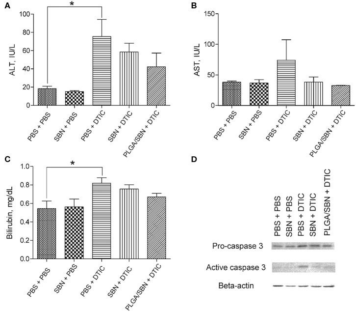
Drug-induced hepatotoxicity is one of the major barriers limiting application of current pharmaceuticals as well as clinical translation of novel and perspective drugs. In this context, numerous hepatoprotective molecules have been proposed to prevent or mitigate drug-induced hepatotoxicity. To date, silibinin (SBN) is a one the most studied hepatoprotective plant-derived agents for prevention/alleviation of drug-induced liver injury. Hepatoprotective mechanisms of SBN include scavenging of free radicals, upregulation of detoxifying enzymes via Nrf2 activation and inhibition of inflammatory activation of resident macrophages. However, low solubility of this phytochemical in water prevents its intravenous administration and constrains its bioavailability and efficacy. Here, we developed SBN-loaded poly(lactic-co-glycolic) acid (PLGA)-based nanoparticles for intravenous administration aiming at mitigation of drug-induced hepatotoxicity. Obtained nanoparticles demonstrated a slow drug release profile in vitro and caused upregulation of antioxidant and phase II enzymes in AML12 hepatocytes including superoxide dismutase 2, glutathione-S-transferase P1, and glutathione-reductase. Intravenous administration of PLGA nanoparticles to mice led to their fast liver accumulation. In vivo analysis of hepatoprotective effects of PLGA/SBN nanoparticles was carried out on melanoma tumor-bearing syngeneic mouse model treated with the antineoplastic drug dacarbazine (DTIC), which often causes severe hepatotoxicity including development of veno-occlusive disease. It was found that PLGA/SBN caused effective induction of detoxifying liver enzymes. Moreover, pre-treatment with PLGA/SBN nanoparticles reduced elevated transaminase and bilirubin levels in blood, caspase 3 activation, and morphological histology changes in liver tissue upon DTIC treatment. Treatment with PLGA/SBN nanoparticles did not interfere with therapeutic efficacy of DTIC.

Keywords: drug combination; drug-induced liver injury; hepatoprotection; melanoma; nanoformulation.


Figures

Figure 1
Figure 1
PLGA/SBN nanoparticle characterization and cytotoxicity. (A) SEM image of PLGA/SBN nanoparticles. Scale bar is 1 μm. (B) In vitro SBN release profile from PLGA/SBN nanoparticles in PBS at 37°C. (C) Viability of AML12 hepatocytes after 24 h of incubation with SBN or PLGA/SBN nanoparticles, determined by MTT assay.
Figure 2
Figure 2
Induction of antioxidant response enzyme expression and hepatoprotective effect by SBN formulations in AML12 hepatocytes. (A) Western blotting analysis of liver enzyme expression in AML12 hepatocytes after treatment with blank PLGA nanoparticles, free SBN and PLGA/SBN nanoparticles. (B) GST activity kinetics in AML12 cells, incubated with 100 μM SBN, measured using CDNB assay. (C) Images of AML12 cells demonstrating antiapoptotic effect of free SBN or PLGA/SBN nanoparticle pre-treatments during incubation with 0.5 mM DTIC. White arrowheads point out single apoptotic cells, whereas white stars denote aggregations of apoptotic cells. Scale bar is 100 μm. (D) Reduction of caspase 3/7 activation in pre-treated with SBN-containing formulations AML12 cells during incubation with DTIC. All values are shown as means ± SD. *P < 0.05, **P < 0.01, ***P < 0.001 (one-way ANOVA followed by a post-hoc Dunnett's t-test) for (B,D).
Figure 3
Figure 3
PLGA nanoparticle biodistribution and upregulation of phase II enzymes by SBN-containing formulations. (A) Analysis of PLGA/rhodamine nanoparticle biodistribution in different tissues of B16F1 tumor-bearing mice upon intravenous administration. All values are shown as means ± SD. (B) Expression levels of phase II and antioxidant enzymes in liver tissue of mice treated with SBN formulations, determined by Western blotting analysis.
Figure 4
Figure 4
The impact of SBN-containing hepatoprotective formulation on anticancer therapeutic effect of DTIC. (A) Images of excised B16F1 tumors from different groups of mice. Scale bar is 5 mm. (B) Kinetics of tumor growth. Pre-treatments with PBS either SBN formulations were performed on the days 3, 6, and 9 after inoculation of cancer cells. Treatments with antineoplastic drug DTIC or PBS (in control groups) were made on the days 4, 7, and 10. All values are shown as means ± SEM. **P < 0.01, ***P < 0.001 (one-way ANOVA followed by a post-hoc Tukey's test).
Figure 5
Figure 5
Hepatoprotective efficacy of SBN formulation pre-treatment. (A) ALT and (B) AST levels in blood serum of tumor-bearing mice after experiment with DTIC therapy. (C) Bilirubin content in blood serum. (D) Pro-caspase 3 and active caspase 3 levels in livers of treated tumor-bearing mice determined by Western blot analysis. All values are shown as means ± SD. *P < 0.05 for (A,C) (one-way ANOVA followed by a post-hoc Dunnett's t-test).
Figure 6
Figure 6
Morphology of liver tissue after chemotherapy with DTIC. Tissue slices of 5 μm thickness were stained with haematoxilyn-eosin. Black arrows denote mononuclear infiltration of sinusoids; black arrowheads denote necrotic foci; yellow arrowheads point to mild cholestasis. Scale bar is 50 μm.

Similar articles

Cited by

References

    1. Abenavoli L., Izzo A. A., Milić N., Cicala C., Santini A., Capasso R. (2018). Milk thistle (Silybum marianum): a concise overview on its chemistry, pharmacological, and nutraceutical uses in liver diseases. Phytother. Res. 32, 2202–2213. 10.1002/ptr.6171 - DOI - PubMed
    1. Au A. Y., Hasenwinkel J. M., Frondoza C. G. (2013). Hepatoprotective effects of S-adenosylmethionine and silybin on canine hepatocytes in vitro. J. Anim. Phys. Anim. Nutr. 97, 331–341. 10.1111/j.1439-0396.2012.01275.x - DOI - PubMed
    1. Bantel H., Lügering A., Poremba C., Lügering N., Held J., Domschke W., et al. . (2001). Caspase activation correlates with the degree of inflammatory liver injury in chronic hepatitis C virus infection. Hepatology 34, 758–767. 10.1053/jhep.2001.28229 - DOI - PubMed
    1. Bektur N. E., Sahin E., Baycu C., Unver G. (2016). Protective effects of silymarin against acetaminophen-induced hepatotoxicity and nephrotoxicity in mice. Toxicol. Ind. Health. 32, 589–600. 10.1177/0748233713502841 - DOI - PubMed
    1. Björnsson E. S. (2016). Hepatotoxicity by drugs: the most common implicated agents. Int. J. Mol. Sci. 17:224. 10.3390/ijms17020224 - DOI - PMC - PubMed