

Comparative Pathogenicity of Duck Hepatitis A Virus-1 Isolates in Experimentally Infected Pekin and Muscovy Ducklings
- PMID: 32671102
- PMCID: PMC7326108
- DOI: 10.3389/fvets.2020.00234
Comparative Pathogenicity of Duck Hepatitis A Virus-1 Isolates in Experimentally Infected Pekin and Muscovy Ducklings
Abstract
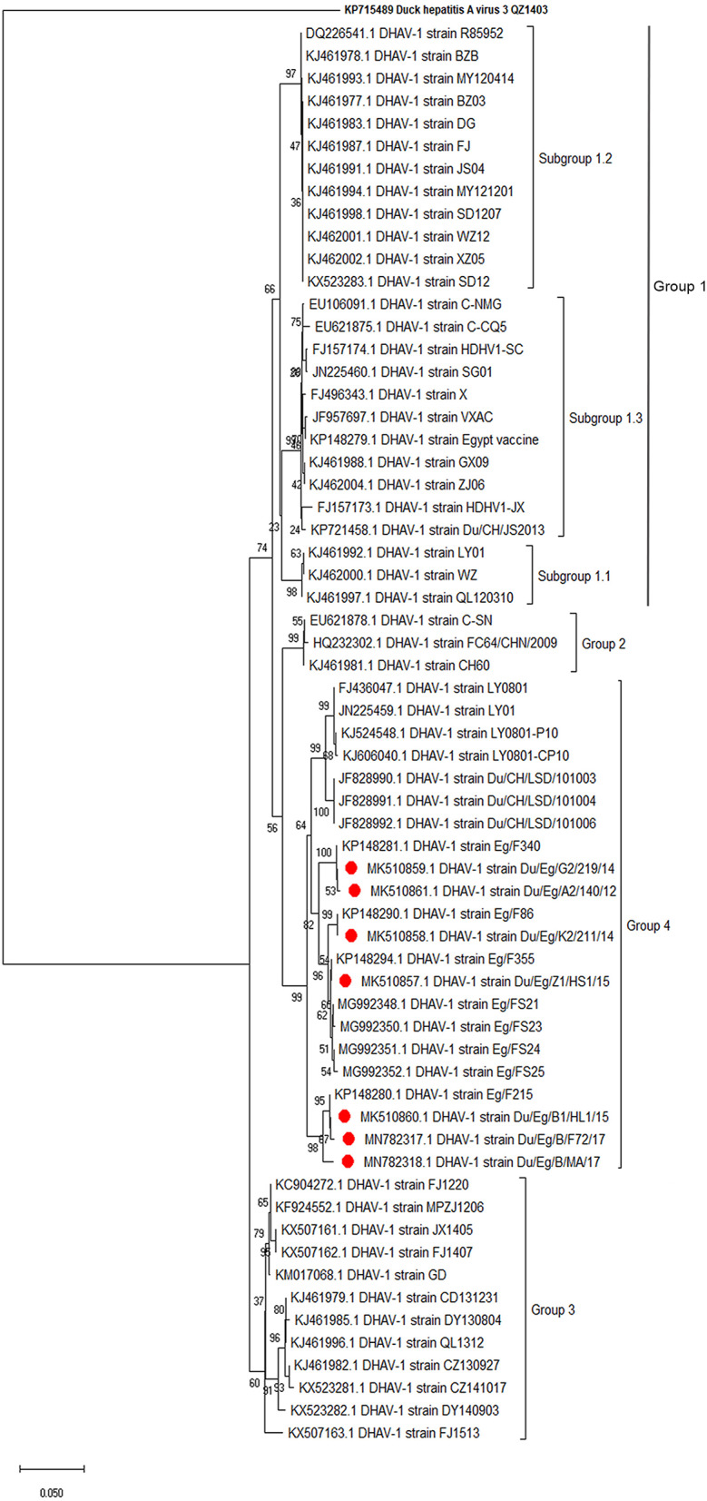
Duck hepatitis virus (DHV) has always been considered one of the threats endangering duck farming in Egypt since the 1960s. In the current study, suspected DHV samples (n = 30) were obtained from commercial Pekin, Mulard (hybrid), and Muscovy duck farms and backyards in Beheira, Alexandria, Gharbia, Kafr El-Sheikh, and Giza provinces between 2012 and 2017. Diseased 3-21-day-old ducklings showed a clinical history of high mortality rates and nervous signs. Samples were screened by RT-PCR targeting the 5'UTR region and VP1 gene. The PCR-confirmed samples (n = 7) were isolated via allantoic route inoculation onto 9-day-old specific-pathogen-free embryonated chicken eggs. Embryos showed stunting, subcutaneous hemorrhages, and liver necrotic greenish-yellow foci. Duck hepatitis A virus-1 (DHAV-1) isolates were genetically analyzed in comparison to other field and vaccine strains. Phylogenetic analyses of the full-length VP1 gene sequences revealed that the obtained DHAV-1 field isolates clustered into genetic group 4 alongside other Egyptian strains isolated during the same period (95.9-99.72% similarity). Amino acid substitutions in the carboxyl-terminal of VP1 (I180T, G184E, D193N, and M213I) were identified in two strains. Also, deletion mutation at I189 was detected in three DHAV-1 strains. Additionally, the two amino acid residues E205 and N235 were common among the isolated strains and other virulent DHAV-1 strains. Two DHAV-1 isolates originated from Pekin source were selected for conducting the comparative pathogenicity testing based on detected point mutations at C-terminus of VP1. We evaluated the pathogenicity of these isolates by investigating clinical signs, mortality rates, and gross pathological and microscopic lesions. The study revealed that experimentally infected Pekin and Muscovy ducklings showed similar clinical signs including squatting down, lateral recumbency, and spasmodic kicking. Muscovy showed milder pathological changes in the liver compared to Pekin ducklings. Histopathological findings supported the gross pathological lesions detected in both breeds. In conclusion, these data provide updated information on the genetic diversity and pathotyping of Egyptian DHAV-1 strains. To the best of our knowledge, this is the first report of comparative pathogenicity of recent DHAV-1 strains in Pekin and Muscovy ducklings in Egypt and the Middle East region.
Keywords: Egypt; duck hepatitis A virus-1 (DHAV-1); duckling; muscovy; pathogenicity; pekin.
Copyright © 2020 Hisham, Ellakany, Selim, Abdalla, Zain El-Abideen, Kilany, Ali and Elbestawy.
Figures


Similar articles
-
Comparative pathogenicity of duck hepatitis A virus genotype 3 in different duck breeds: Implications of the diagnosis and prevention of duck viral hepatitis.Comp Immunol Microbiol Infect Dis. 2024 Nov;114:102256. doi: 10.1016/j.cimid.2024.102256. Epub 2024 Oct 13. Comp Immunol Microbiol Infect Dis. 2024. PMID: 39437532
-
Outbreaks of Duck Hepatitis A Virus in Egyptian Duckling Flocks.Avian Dis. 2019 Mar 1;63(1):68-74. doi: 10.1637/11975-092118-Reg.1. Avian Dis. 2019. PMID: 31251521
-
Epidemiology and molecular characterisation of duck hepatitis A virus from different duck breeds in Egypt.Vet Microbiol. 2015 Jun 12;177(3-4):347-52. doi: 10.1016/j.vetmic.2015.03.020. Epub 2015 Mar 27. Vet Microbiol. 2015. PMID: 25862279
-
Hepatitis Virus Infections in Poultry.Avian Dis. 2016 Sep;60(3):576-88. doi: 10.1637/11229-070515-Review.1. Avian Dis. 2016. PMID: 27610716 Review.
-
Duck hepatitis A virus prevalence in mainland China between 2009 and 2021: A systematic review and meta-analysis.Prev Vet Med. 2022 Nov;208:105730. doi: 10.1016/j.prevetmed.2022.105730. Epub 2022 Jul 30. Prev Vet Med. 2022. PMID: 35964373
Cited by
-
Insights into the Genetic Evolution of Duck Hepatitis A Virus in Egypt.Animals (Basel). 2021 Sep 19;11(9):2741. doi: 10.3390/ani11092741. Animals (Basel). 2021. PMID: 34573707 Free PMC article.
-
A comprehensive pathological and molecular investigation of viral co-infections in ducks in Egypt.Front Microbiol. 2025 May 8;16:1522669. doi: 10.3389/fmicb.2025.1522669. eCollection 2025. Front Microbiol. 2025. PMID: 40406342 Free PMC article.
-
Genomic Epidemiology and Evolution of Duck Hepatitis A Virus.Viruses. 2021 Aug 11;13(8):1592. doi: 10.3390/v13081592. Viruses. 2021. PMID: 34452457 Free PMC article.
-
One-Step Multiplex Real-Time Fluorescent Quantitative Reverse Transcription PCR for Simultaneous Detection of Four Waterfowl Viruses.Microorganisms. 2024 Nov 25;12(12):2423. doi: 10.3390/microorganisms12122423. Microorganisms. 2024. PMID: 39770626 Free PMC article.
-
The Impact of Genetic Variation on Duck Hepatitis A Virus (DHAV) Vaccine Efficacy: A Comparative Study of DHAV-1 and DHAV-3 Against Emerging Variant Strains.Vaccines (Basel). 2024 Dec 16;12(12):1416. doi: 10.3390/vaccines12121416. Vaccines (Basel). 2024. PMID: 39772077 Free PMC article.
References
-
- Tsai HJ. Duck hepatitis. In: Swayne DE, editors. Diseases of Poultry. Hoboken, NJ: Wiley-Blackwell; (2020). p. 450–9. 10.1002/9781119371199 - DOI
LinkOut - more resources
Full Text Sources