. 2020 Jul 8;6(7):e04392.
doi: 10.1016/j.heliyon.2020.e04392. eCollection 2020 Jul.

Alpha glucosidase inhibition activity of phenolic fraction from Simarouba glauca: An in-vitro, in-silico and kinetic study

Affiliations

Alpha glucosidase inhibition activity of phenolic fraction from Simarouba glauca: An in-vitro, in-silico and kinetic study

Kirana P Mugaranja et al. Heliyon. .

Abstract

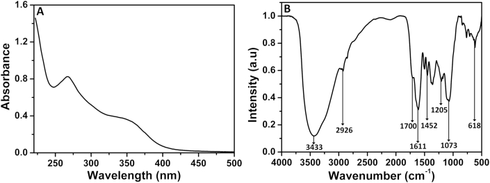
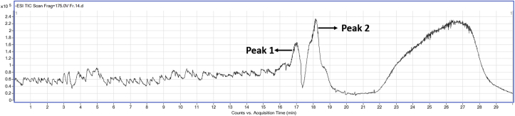
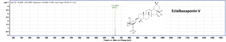
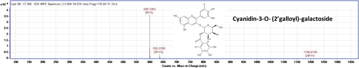
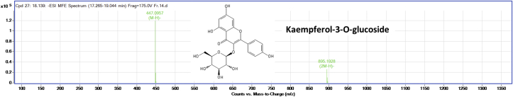
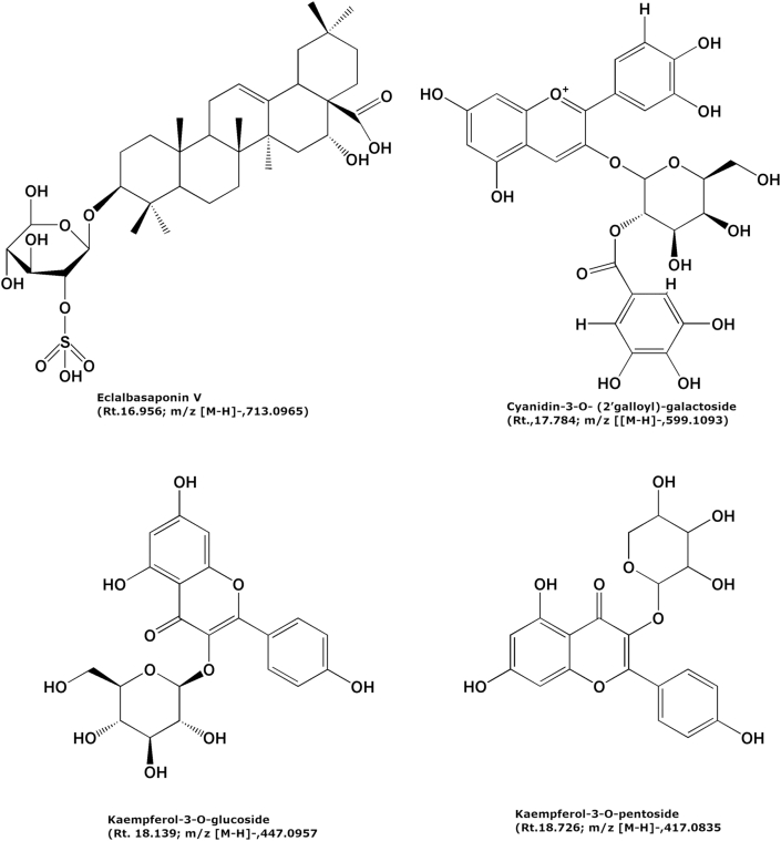
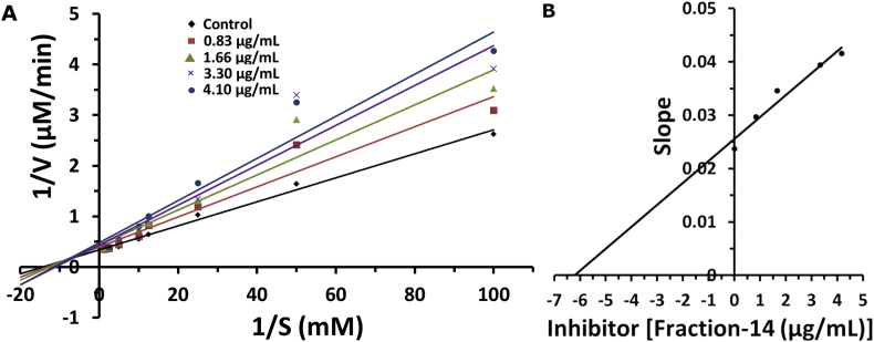
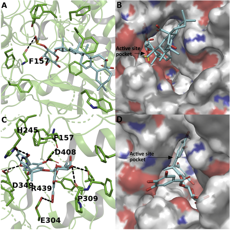
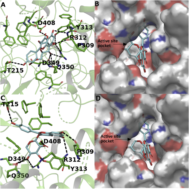
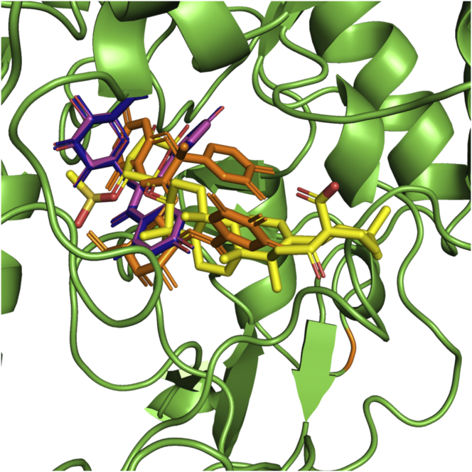
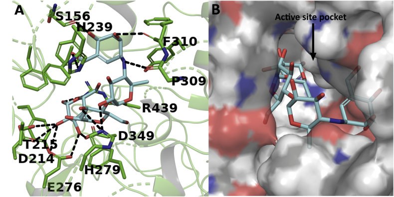
A phenolic rich fraction purified from Simarouba glauca leaves was effective in alpha glucosidase inhibition. The purified fraction named 'fraction-14' had shown significant inhibition of yeast alpha glucosidase enzyme activity (IC50 = 2.4 ± 0.4 μg/mL) when compared to anti-diabetic drug acarbose (IC50 = 2450 ± 24 μg/mL). The purified fraction also had reasonable DPPH (IC50 = 14.4 ± 0.1 μg/mL) and ABTS (IC50 = 7.6 ± 0.5 μg/mL) free radical scavenging activity when compared to the standard ascorbic acid. The LC-MS analysis of bioactive 'fraction-14' revealed four compounds, eclalbasaponin-v (1), cyanidin-3-O-(2'galloyl)-galactoside (2), kaempferol-3-O-glucoside (3) and kaempferol-3-O-pentoside (4) for the first time in S. glauca in this study. The kinetic study of the 'fraction-14' indicates a mixed type of inhibition on the alpha glucosidase enzyme with K i , 6.2 μg/mL. Docking studies showed promising binding energy for the compounds 2 (-7.769 kJ/mol), 3 (-7.04 kJ/mol) and 4 (-7.127 kJ/mol) against yeast alpha glucosidase which was better than acarbose (-6.867 kJ/mol). In conclusion, the phenolic rich fraction from S. glauca possessing good in-vitro antioxidant property and alpha glucosidase enzyme inhibition potential along with mixed inhibition kinetics. Also, better binding energy of compounds (1, 2 & 3) appears to contain potential lead-molecule for antidiabetic therapy.

Keywords: Agriculture; Alpha glucosidas; Anti-diabetic; Enzyme kinetics; Food chemistry; Food science; Hypoglycaemic; Medicinal plant; Molecular docking.


Figures

Figure 1
Figure 1
Silica gel column purified phenolic rich ‘fraction-14’. Out of 16 fractions colleced from the silica gel column chromatography of ethanol extract from S. glauca leaf, the ‘fraction-14’ had high phenolic content and found to be highly active against alpha glucosidase enzyme.
Figure 2
Figure 2
UV-Visible spectroscopic analysis (A) and FTIR spectra (B) of column purified ‘fraction-14’ of ethanol extract from S. glauca leaf. For UV-Visible spectroscopy, the sample was dissolved in Milli-Q water at 0.25 mg/mL concentration. For FTIR, the sample was mixed with KBr at a ratio of 1:100, ground into a fine powder and a thin pellet was made.
Figure 3
Figure 3
ESI-TIC spectra of column purified ‘fraction-14’. The LC-MS peaks at retention time between 16.4-18.8 min which has high intensity was analysed for mass spectroscopy.
Figure 4
Figure 4
Mass spectra of eclalbasaponin V (compound 1). The m/z value was obtained in negative ionization mode and compared with m/z value of previously identified compounds in literature.
Figure 5
Figure 5
Mass spectra of cyanidin-3-O- (2′galloyl)-galactoside (compound 2). The m/z value was obtained in negative ionization mode and compared with m/z value of previously identified compounds in literature.
Figure 6
Figure 6
Mass spectra of kaempferol-3-O-glucoside (compound 3). The m/z value was obtained in negative ionization mode and compared with m/z value of previously identified compounds in literature.
Figure 7
Figure 7
Mass spectra of kaempferol-3-O-pentoside (compound 4). The m/z value was obtained in negative ionization mode and compared with m/z value of previously identified compounds in literature.
Figure 8
Figure 8
Chemical structures of tentatively identified compounds from column purified ‘fraction-14’ of ethanol extract from S. glauca leaf using LC-MS analysis. The compounds were identified based on the comparison of m/z values with m/z values of previously identified compounds in literature.
Figure 9
Figure 9
Lineweaver- Burk plots (A) and Secondary plot of slope versus inhibitor (‘fraction-14’) (B). The inhibitor concentrations were used as 0.83, 1.66, 3.32 and 4.15 μg/mL. Alpha glucosidase concentration was 0.02 units/mL. Substrate, PNPG concentrations were 0.01, 0.02, 0.04, 0.08, 0.1, 0.2, 0.4 and 0.8 mM.
Figure 10
Figure 10
Surface representation of modelled structure of the yeast alpha glucosidase. The structure was generated using RaptorX software. The active site of the enzyme is represented in a yellow-colored patch where the docked compounds interacts with enzyme which involves both hydrogen bonds and hydrophobic interactions.
Figure 11
Figure 11
Ramachandran plot for homology model of yeast alpha glucosidase. The homology model was built using RaptorX software. The model was validated and authenticated by Ramachandran plot using PROCHECK software.
Figure 12
Figure 12
Docking study of compounds 1 and 2 with yeast alpha glucosidase. Interaction of compound 1 in stick representation (A) and surface representation form (B); and interaction of compound 2 in stick representation (C) and surface representation form (D) with active site residues of alpha glucosidase. The compounds were perfectly stacked inside the active site pocket of the enzyme with several hydrogen bonds and hydrophobic interactions. The hydrogen bond interactions were represented in black coloured broken lines.
Figure 13
Figure 13
Docking study of compounds 3 and 4 with yeast alpha glucosidase. Interaction of compound 3 in stick representation (A) and surface representation form (B); and interaction of compound 4 in stick representation (C) and surface representation form (D) with active site residues of alpha glucosidase. The compounds were perfectly stacked inside the active site pocket of the enzyme with several hydrogen bonds and hydrophobic interactions. The hydrogen bond interactions were represented in black coloured broken lines.
Figure 14
Figure 14
Superimposition of the docked compounds in the active site of the enzyme. Compounds 1, 2, 3 and 4 were represented in yellow, orange, blue and purple coloured sticks respectively.
Figure 15
Figure 15
Docking study of acarbose with yeast alpha glucosidase. (A) Stick representation and (B) Surface representation. The hydrogen bond interactions were represented in black coloured broken lines.

References

    1. Abu-Reidah I.M., Ali-Shtayeh M.S., Jamous R.M., Arráez-Román D., Segura-Carretero A. HPLC–DAD–ESI-MS/MS screening of bioactive components from Rhus coriaria L.(Sumac) fruits. Food Chem. 2015;166:179–191. - PubMed
    1. Alam M.A., Zaidul I., Ghafoor K., Sahena F., Hakim M., Rafii M., Abir H., Bostanudin M., Perumal V., Khatib A. In vitro antioxidant and, α-glucosidase inhibitory activities and comprehensive metabolite profiling of methanol extract and its fractions from Clinacanthus nutans. BMC Compl. Alternative Med. 2017;17(181):1–10. - PMC - PubMed
    1. Alves I.A., Miranda H.M., Soares L.A., Randau K.P. Simaroubaceae family: botany, chemical composition and biological activities. Revista Brasileira de Farmacognosia. 2014;24(4):481–501.
    1. Amarowicz R., Pegg R., Rahimi-Moghaddam P., Barl B., Weil J. Free-radical scavenging capacity and antioxidant activity of selected plant species from the Canadian prairies. Food Chem. 2004;84(4):551–562.
    1. Blando F., Calabriso N., Berland H., Maiorano G., Gerardi C., Carluccio M.A., Andersen Ø.M. Radical scavenging and anti-inflammatory activities of representative anthocyanin groupings from pigment-rich fruits and vegetables. Int. J. Mol. Sci. 2018;19(169):1–15. - PMC - PubMed

LinkOut - more resources