Case Reports
. 2020 Jul 3:19:100798.
doi: 10.1016/j.ajoc.2020.100798. eCollection 2020 Sep.

Epiphora and unrecognized paranasal sinuses pathology

Affiliations
Case Reports

Epiphora and unrecognized paranasal sinuses pathology

Filippo Confalonieri et al. Am J Ophthalmol Case Rep. .

Abstract

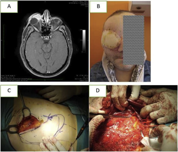
Purpose: to report five patients all presenting with persistent unilateral epiphora as a sign of unexpected and rare lesions causing Secondary Acquired Nasolacrimal Duct Obstruction (SANDO) and the risks associated to an incomplete diagnostic work-up.

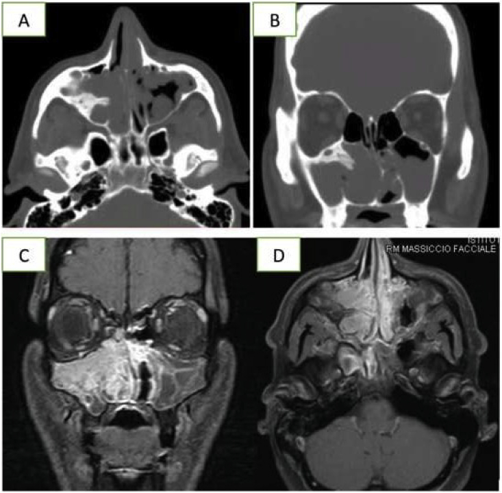
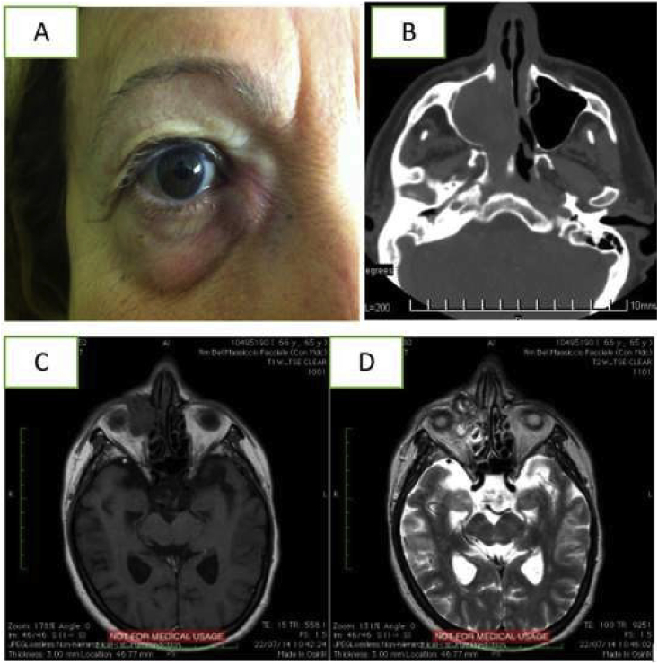
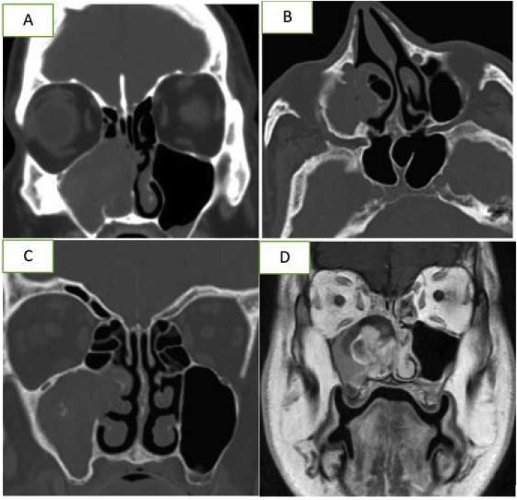
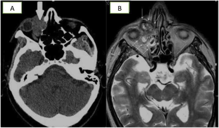
Observations: the cases presented are: (1) Fungus ball, (2) Pyogenic granuloma, (3) Sinonasal inverted papilloma (4) Sinonasal inverted papilloma with synchronous squamous cell carcinoma, (5) Squamous cell carcinoma of the lacrimal sac.

Conclusions and importance: masses are uncommon but not a rare cause of nasolacrimal duct obstruction. Surgical teams performing large numbers of dacryocystorhinostomies should be aware of such pathology and perform a systematic multidisciplinary approach.

Keywords: Fungus ball; Lacrimal sac tumors; Pyogenic granuloma; Sinonasal inverted papilloma; Squamous cell carcinoma; Tearing.


Conflict of interest statement

The following authors have no financial disclosures: ADM, FC, RP, LB, LM.

Figures

Fig. 1
Fig. 1
Case 1 (A–B): heterogeneous CT signaling due to areas of high attenuation caused by ferromagnetic elements (iron-like pattern) with middle turbinate pneumatization (concha bullosa). (C–D): MRI scan of the same patient.
Fig. 2
Fig. 2
Case 2 (A): External appearance of patient. CT (B) and Gadolinium MRI (C, D) showing the radiological pattern of the bulky tumor centered on the inferior turbinate and the lateral nasal wall with involvement of Hasner's valve. The high vascularity is responsible for signal voids. The medial wall of the maxillary sinus and the nasolacrimal duct manifest erosion due to the compression but there is no radiological sign of secondary to bone infiltration.
Fig. 3
Fig. 3
Case 3 (A–B): CT scan showing the erosion of the middle wall of maxillary sinus and the hyperostosis of the inferior orbital wall as only peculiar sign. (C–D): MRI pointing out the characteristic features of inverted papilloma, including the convoluted pattern on contrast-enhanced T1 and T2-weighted images. This characteristic aspect is related to the alternating hypointense and hyperintense signal within the mass.
Fig. 4
Fig. 4
Case 4 (A) Non enhanced axial CT shows bone erosion of the papyracea lamina of ethmoid bone. (B) Axial T2 weighted sequence shows an extraconal and intraconal mass (white arrows) in the right orbit, with non-homogeneous hyperintensity ed irregular margins. The lesion involves the nasolacrimal canal, the papyracea lamina and the anterior ethmoid cells.
Fig. 5
Fig. 5
Case 5 (A) T1-weighted MRI showing a mass involving the lacrimal sac and extending to the orbit. (B) 1-month follow-up appearance. (C–D) The orbital cavity was filled with a pedunculated flap of rectus abdominis through anastomosis with internal jugular vein and facial artery.

Similar articles

Cited by

References

    1. Barry K., Jackson J., Williams K. The tearing patient. Elsevier Disease-a-Month. March 2017;63(3):68–71. - PubMed
    1. Nemet Arie Y. The etiology of epiphora: a multifactorial issue. Semin Ophthalmol. 2016;31(3):275–279. doi: 10.3109/08820538.2014.962163. - DOI - PubMed
    1. Paulsen F. New insights into the pathophysiology of primary acquired dacryostenosis. Ophthalmology. 2001;108(12):2329–2336. doi: 10.1016/S0161-6420(01)00946-0. - DOI - PubMed
    1. Yazici H., Bulbul E., Yazici A. Primary acquired nasolacrimal duct obstruction: is it really related to paranasal abnormalities? Surg Radiol Anat. 2015;37:579–584. doi: 10.1007/s00276-014-1391-6. - DOI - PubMed
    1. Linberg J.V., McCormick S.A. Primary acquired nasolacrimal duct obstruction. A clinicopathologic report and biopsy technique. Ophthalmology. 1986;93:1055–1063. - PubMed

Publication types

LinkOut - more resources