Comment
. 2020 Sep;11(9):680-687.
doi: 10.1007/s13238-020-00752-4.

Single-cell analysis reveals bronchoalveolar epithelial dysfunction in COVID-19 patients

Affiliations
Comment

Single-cell analysis reveals bronchoalveolar epithelial dysfunction in COVID-19 patients

Jiangping He et al. Protein Cell. 2020 Sep.
No abstract available


Figures

Figure 1
Figure 1
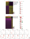
Mucin hypersecretion from Club cells upon SARS-CoV-2 infection. (A) (Upper) UMAP plots visualizing single-cell RNA-seq data of 4,237 BALF single cells from COVID-19 patients and 6,100 lung single cells from 8 healthy control (HC). (Lower) Differences in cellular composition among HC (green, left) and BALF (red, right). (B) Heatmap showing the average expression levels per cluster of the differentially expressed markers in each cluster. (C) UMAP depicting the expression of MUC5AC in HC and COVID-19 patients. (D) Heatmap depicting the differential expressed genes between HC and COVID-19 patients in Club cells. Selected genes are labeled in the right. All genes are significant differential expressed with fold change >1.5 and P-value < 0.01; P-value was from the Wilcoxon test. (E) Violin plots showing the differential expression of MUC genes between HC and COVID-19 patients. *** represents P-value < 0.001, P-value was from Wilcoxon test. (F) Gene ontology (GO) analysis for the differential expressed genes from panel (D). (G) Motif and TF enrichment for the differentially expressed genes from panel (D). The motif ranking database hg38__refseq-r80__10kb_up_and_down_tss was used for enrichment and motifs were group by the family of the corresponding transcription factors. (H) Heatmap depicting the differential expressed genes in Ciliated cells between HC and COVID-19 patients. (I) Violin plots showing the expression of cilium formation related genes between HC and COVID-19 patients. (J) GO analysis for the differential expressed genes from panel (H)
Figure 1
Figure 1
Mucin hypersecretion from Club cells upon SARS-CoV-2 infection. (A) (Upper) UMAP plots visualizing single-cell RNA-seq data of 4,237 BALF single cells from COVID-19 patients and 6,100 lung single cells from 8 healthy control (HC). (Lower) Differences in cellular composition among HC (green, left) and BALF (red, right). (B) Heatmap showing the average expression levels per cluster of the differentially expressed markers in each cluster. (C) UMAP depicting the expression of MUC5AC in HC and COVID-19 patients. (D) Heatmap depicting the differential expressed genes between HC and COVID-19 patients in Club cells. Selected genes are labeled in the right. All genes are significant differential expressed with fold change >1.5 and P-value < 0.01; P-value was from the Wilcoxon test. (E) Violin plots showing the differential expression of MUC genes between HC and COVID-19 patients. *** represents P-value < 0.001, P-value was from Wilcoxon test. (F) Gene ontology (GO) analysis for the differential expressed genes from panel (D). (G) Motif and TF enrichment for the differentially expressed genes from panel (D). The motif ranking database hg38__refseq-r80__10kb_up_and_down_tss was used for enrichment and motifs were group by the family of the corresponding transcription factors. (H) Heatmap depicting the differential expressed genes in Ciliated cells between HC and COVID-19 patients. (I) Violin plots showing the expression of cilium formation related genes between HC and COVID-19 patients. (J) GO analysis for the differential expressed genes from panel (H)
Figure 2
Figure 2
Differential expressing genes reveals macrophages and monocytes/neutrophils anti-viral response. (A) Heatmap showing the differential expressed genes between HC and COVID-19 patients in macrophages, selected genes are indicated in the right. (B) Violin plots depicting the selected differential expressed genes between HC and COVID-19 patients in macrophages. (C) Heatmap showing the differential expressed genes between HC and COVID-19 patients in monocytes/neutrophils. (D) Violin plots depicting the selected differential expressed genes between HC and COVID-19 patients in monocytes/neutrophils. (E) UMAP showing the expression of selected genes between HC and COVID-19 patients. (F) Violin plots showing the expression of FCN1 in macrophages (upper) or monocytes/neutrophils (lower). (G) Schematic diagram of the normal alveolus (left) and the severe COVID-19 alveolus (right)
Figure 2
Figure 2
Differential expressing genes reveals macrophages and monocytes/neutrophils anti-viral response. (A) Heatmap showing the differential expressed genes between HC and COVID-19 patients in macrophages, selected genes are indicated in the right. (B) Violin plots depicting the selected differential expressed genes between HC and COVID-19 patients in macrophages. (C) Heatmap showing the differential expressed genes between HC and COVID-19 patients in monocytes/neutrophils. (D) Violin plots depicting the selected differential expressed genes between HC and COVID-19 patients in monocytes/neutrophils. (E) UMAP showing the expression of selected genes between HC and COVID-19 patients. (F) Violin plots showing the expression of FCN1 in macrophages (upper) or monocytes/neutrophils (lower). (G) Schematic diagram of the normal alveolus (left) and the severe COVID-19 alveolus (right)

Comment on

References

    1. Bian X-W, Team TC-P (2020) Autopsy of COVID-19 victims in China. National Science Review. - PMC - PubMed
    1. Bustamante-Marin XM, Ostrowski LE. Cilia and mucociliary clearance. Cold Spring Harb Perspect Biol. 2017;9:28241. doi: 10.1101/cshperspect.a028241. - DOI - PMC - PubMed
    1. Cavalli G, De Luca G, Campochiaro C, Della-Torre E, Ripa M, Canetti D, Oltolini C, Castiglioni B, Tassan Din C, Boffini N, et al. Interleukin-1 blockade with high-dose anakinra in patients with COVID-19, acute respiratory distress syndrome, and hyperinflammation: a retrospective cohort study. Lancet Rheumatol. 2020;2:e325–e331. doi: 10.1016/S2665-9913(20)30127-2. - DOI - PMC - PubMed
    1. Chen AJ, Dong J, Yuan XH, Bo H, Li SZ, Wang C, Duan ZJ, Zheng LS. Anti-H7N9 avian influenza A virus activity of interferon in pseudostratified human airway epithelium cell cultures. Virol J. 2019;16:44. doi: 10.1186/s12985-019-1146-4. - DOI - PMC - PubMed
    1. Chivukula RR, Montoro DT, Leung HM, Yang J, Shamseldin HE, Taylor MS, Dougherty GW, Zariwala MA, Carson J, Daniels MLA, et al. A human ciliopathy reveals essential functions for NEK10 in airway mucociliary clearance. Nat Med. 2020;26:244–251. doi: 10.1038/s41591-019-0730-x. - DOI - PMC - PubMed