

Performance of the Rule of 5 for Detecting Glaucoma Progression between Visits with OCT
- PMID: 32672674
- PMCID: PMC7375168
- DOI: 10.1016/j.ogla.2019.05.003
Performance of the Rule of 5 for Detecting Glaucoma Progression between Visits with OCT
Abstract
Purpose: To evaluate whether loss of 5 μm or more in global retinal nerve fiber layer (RNFL) thickness on spectral-domain (SD) between 2 consecutive visits is specific for glaucoma progression.
Design: Prospective cohort.
Participants: Ninety-two eyes of 49 control participants and 300 eyes of 210 glaucoma patients.
Methods: Patients completed at least 5 standard automated perimetry and SD OCT examinations at 6-month intervals over at least 2 years. Eyes were categorized as progressing from glaucoma if the average RNFL declined by 5 μm or more between 2 consecutive visits. The false-positive proportion was estimated by 2 methods: (1) 5-μm or more loss in control participants and (2) 5-μm or more gain in glaucoma. The false-positive proportion was subtracted from the cumulative proportion of eyes categorized with glaucoma progression to estimate the true progression prevalence.
Main outcome measures: False-positive and true progression prevalence of patients with glaucoma detected as progressing on SD OCT.
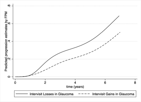
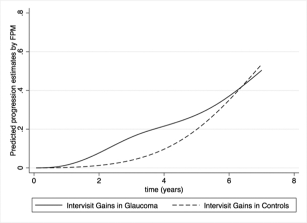
Results: After 5 years of semiannual testing, the cumulative proportion of false-positive results based on 5-μm or more RNFL losses between visits was 24.8% in the control participants. Although 40.6% of glaucoma eyes were diagnosed with progression at 5 years, only 15.8% would have been considered to show true progression based on the expected false-positive ratio from the control participants (i.e., 40.6%-24.8%). The cumulative proportion of an intervisit gain of 5 μm or more at 5 years was 27.4% in glaucoma eyes, suggesting that only 13.2% of eyes with glaucoma truly had progressed (i.e., 40.6%-27.4%).
Conclusions: Loss of 5 μm or more in average RNFL thickness between consecutive SD OCT tests is not specific for glaucoma progression. Application of this intervisit rule of 5 can result in a high cumulative proportion of false-positive results over time, which could lead to unnecessary interventions in patients whose disease is stable. More specific diagnostic criteria are needed to help clinicians determine whether patients with glaucoma are progressing so that therapy escalation is both timely and appropriate.
Copyright © 2019 American Academy of Ophthalmology. Published by Elsevier Inc. All rights reserved.
Conflict of interest statement
Conflict of Interest:
ACT: none. AAJ: none. FAM: Alcon Laboratories (C, F, R), Allergan (C, F), Bausch&Lomb (F), Carl Zeiss Meditec (C, F, R), Heidelberg Engineering (F), Merck (F), nGoggle Inc. (F), Sensimed (C), Topcon (C), Reichert (C, R).
Figures

References
-
- Medeiros FA, Zangwill LM, Alencar LM, et al. Rates of progressive retinal nerve fiber layer loss in glaucoma measured by scanning laser polarimetry. Am J Ophthalmol. 2010;149(6):908–15. - PubMed
-
- Chauhan BC, Hutchison DM, Artes PH, et al. Optic disc progression in glaucoma: comparison of confocal scanning laser tomography to optic disc photographs in a prospective study. Invest Ophthalmol Vis Sci. 2009;50(4):1682–91. - PubMed
Publication types
MeSH terms
Grants and funding
LinkOut - more resources
Full Text Sources
Medical
