. 2020 Jul 16;15(7):e0236199.
doi: 10.1371/journal.pone.0236199. eCollection 2020.

Ultraviolet A light effectively reduces bacteria and viruses including coronavirus

Affiliations

Ultraviolet A light effectively reduces bacteria and viruses including coronavirus

Ali Rezaie et al. PLoS One. .

Erratum in

Abstract

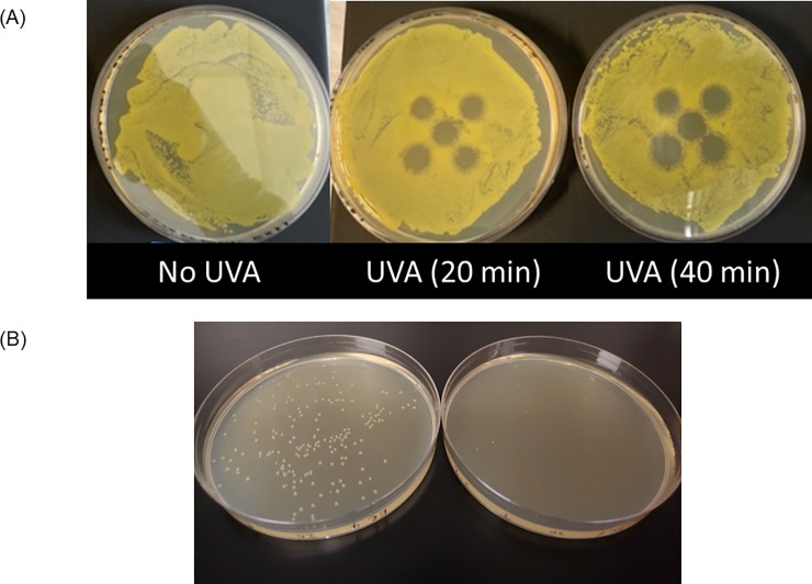
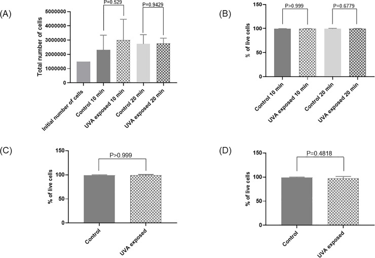
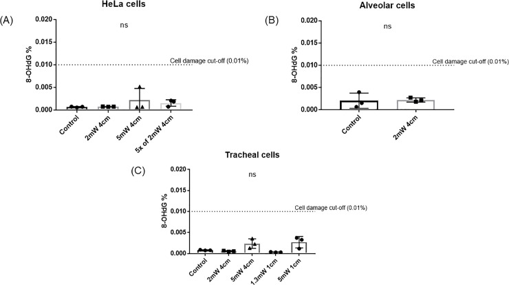
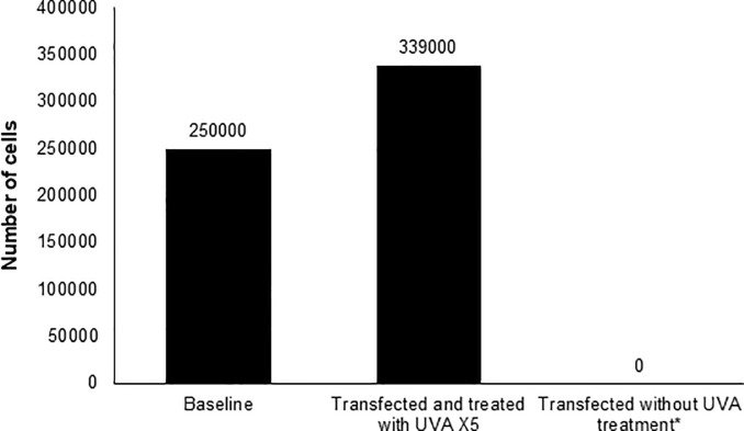
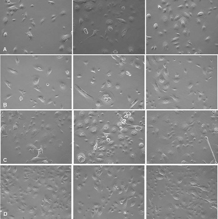
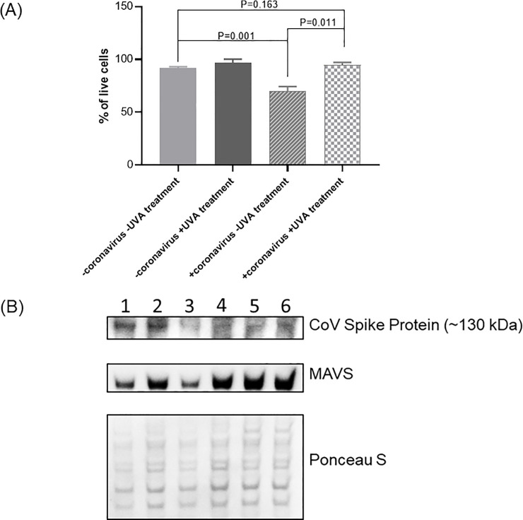
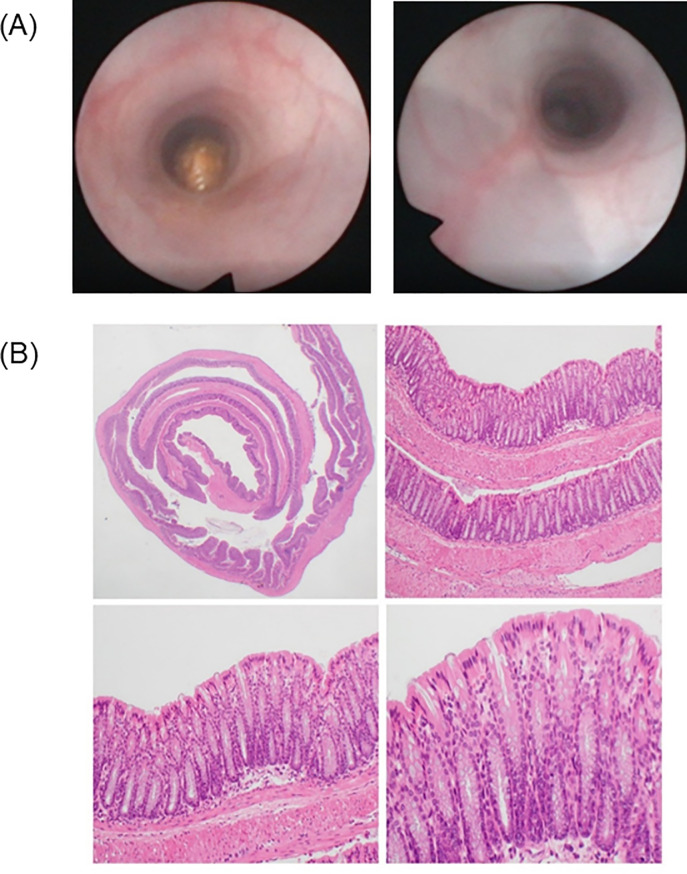
Antimicrobial-resistant and novel pathogens continue to emerge, outpacing efforts to contain and treat them. Therefore, there is a crucial need for safe and effective therapies. Ultraviolet-A (UVA) phototherapy is FDA-approved for several dermatological diseases but not for internal applications. We investigated UVA effects on human cells in vitro, mouse colonic tissue in vivo, and UVA efficacy against bacteria, yeast, coxsackievirus group B and coronavirus-229E. Several pathogens and virally transfected human cells were exposed to a series of specific UVA exposure regimens. HeLa, alveolar and primary human tracheal epithelial cell viability was assessed after UVA exposure, and 8-Oxo-2'-deoxyguanosine was measured as an oxidative DNA damage marker. Furthermore, wild-type mice were exposed to intracolonic UVA as an in vivo model to assess safety of internal UVA exposure. Controlled UVA exposure yielded significant reductions in Pseudomonas aeruginosa, Klebsiella pneumoniae, Escherichia coli, Enterococcus faecalis, Clostridioides difficile, Streptococcus pyogenes, Staphylococcus epidermidis, Proteus mirabilis and Candida albicans. UVA-treated coxsackievirus-transfected HeLa cells exhibited significantly increased cell survival compared to controls. UVA-treated coronavirus-229E-transfected tracheal cells exhibited significant coronavirus spike protein reduction, increased mitochondrial antiviral-signaling protein and decreased coronavirus-229E-induced cell death. Specific controlled UVA exposure had no significant effect on growth or 8-Oxo-2'-deoxyguanosine levels in three types of human cells. Single or repeated in vivo intraluminal UVA exposure produced no discernible endoscopic, histologic or dysplastic changes in mice. These findings suggest that, under specific conditions, UVA reduces various pathogens including coronavirus-229E, and may provide a safe and effective treatment for infectious diseases of internal viscera. Clinical studies are warranted to further elucidate the safety and efficacy of UVA in humans.


Conflict of interest statement

Cedars-Sinai Medical Center has a licensing agreement with Aytu BioSciences. Cedars-Sinai has a patent on internal UV therapy, inventors: AR, MP, GM, RM and GL. SS is an employee of Australian Clinical Labs. The authors declare no other financial conflicts of interest. This does not alter our adherence to PLOS ONE policies on sharing data and materials.

Figures

Fig 1
Fig 1
(A) Effect of narrow-band UVA exposure with intensity of 2000μW/cm2 at the E. coli culture plate. An array of five LEDs was placed 1 cm above the plate, and plates were exposed for 20 and 40 minutes. (B) Effects of UVA treatment of E. coli liquid cultures when subsequently plated. Liquid E. coli cultures were treated with 3000 W/cm2 for 20 minutes (right) or left untreated as controls (left), then plated on solid medium. There is a notable decrease in the number and size of E. coli colonies following UVA treatment.
Fig 2
Fig 2. Effects of UVA treatment on cell growth and viability.
(A) Effect of 10- and 20-minutes narrow-band (NB) UVA exposure (2000 μW/cm2) on total number of HeLa cells (N = 3). (B) Effect of 10- and 20-minutes NB UVA exposure (2000 μW/cm2) on percentage of viable HeLa cells (N = 3). (C) Effect of 20 minutes higher dose NB UVA exposure (5000 μW/cm2) on percentage of viable HeLa cells (N = 3). (D) Effect of 20 minutes UVA exposure (2000 μW/cm2) on percentage of viable alveolar cells (N = 3). Bars represent the mean value of the total number of cells (A) and percentage of live cells (B, C and D) for controls not exposed to UVA and cells exposed to UVA.
Fig 3
Fig 3
8-OHdG levels after exposure to NB-UVA at various intensities at the cell plate on A) HeLa cells, B) alveolar cells and C) ciliated tracheal epithelial cells.
Fig 4
Fig 4. Effect of repeated UVA treatments on the number of adherent HeLa cells post-transfection with Coxsackievirus at 72 hours.
*The number of adherent cells at 48 hours were below the limit of detection of the automated cell counter used (Biorad T20), and no adherent cells were observed at 72 hours.
Fig 5
Fig 5
Effect of UVA treatment on coronavirus 229E infection of HTeC cells at A) 16 hours, B) 36 hours, C) 72 hours and D) 96 hours post transfection (20x phase-contrast images). Left panels: Uninfected, untreated control cells. Middle panels: cells transfected with coronavirus 229E. Right panels: cells transfected with UVA-treated coronavirus 229E and then treated with UVA. Cells transfected with coronavirus 229E exhibit increasing vacuolation and cell death over time, resulting in decreased cell density. In contrast, transfected and UVA-treated cells remain viable and exhibit similar morphology to controls.
Fig 6
Fig 6
A) Viability of ciliated tracheal epithelial cells depending on transfection with coronavirus 229E and treatment with UVA light after 96 hours. Transfected ciliated tracheal epithelial cells showed 25% ±1.84 more viability when treated with NB UVA. B) Intracellular detection of coronavirus 229E in ciliated tracheal epithelial cells treated with NB-UVA light at 96 hours and levels of Mitochondrial antiviral-signaling protein (MAVS). Column 1, 2, and 3 represent cells transfected with CoV-229E; column 4, 5 and 6 –cells transfected with CoV-229E and treated with NB-UVA. Ponceau S Stain was used to locate overall protein bands to check the amount of protein loaded on the gel.
Fig 7
Fig 7
A) Colonoscopic images before and 72 hours after repeated UVA treatment in mice. B) Full-thickness histological examination of the post-mortem colon in BB-UVA-treated mice at various magnifications. Clockwise from top to left, H&E microscopic examinations at 12.5X, 100X, 200X and 400X magnification. There is no evidence of endoscopic or histologic abnormalities.

References

    1. Huang C, Wang Y, Li X, Ren L, Zhao J, Hu Y, et al. Clinical features of patients infected with 2019 novel coronavirus in Wuhan, China. Lancet. 2020;395(10223):497–506. Epub 2020/01/28. 10.1016/S0140-6736(20)30183-5 . - DOI - PMC - PubMed
    1. World Health Organization. Ultraviolet radiation (UV) [cited 2020]. Available from: https://www.who.int/uv/uv_and_health/en/#:~:text=The%20UV%20region%20cov....
    1. World Health Organization. Protecting Workers from Ultraviolet Radiation. World Health Organization; 2007.
    1. Reed NG. The history of ultraviolet germicidal irradiation for air disinfection. Public Health Rep. 2010;125(1):15–27. Epub 2010/04/21. 10.1177/003335491012500105 - DOI - PMC - PubMed
    1. Roy S. Impact of UV Radiation on Genome Stability and Human Health. Adv Exp Med Biol. 2017;996:207–19. Epub 2017/11/11. 10.1007/978-3-319-56017-5_17 . - DOI - PubMed

Publication types

MeSH terms