. 2020 Jul 16;16(7):e1008654.
doi: 10.1371/journal.ppat.1008654. eCollection 2020 Jul.

Mutant prion proteins increase calcium permeability of AMPA receptors, exacerbating excitotoxicity

Affiliations

Mutant prion proteins increase calcium permeability of AMPA receptors, exacerbating excitotoxicity

Elsa Ghirardini et al. PLoS Pathog. .

Erratum in

Abstract

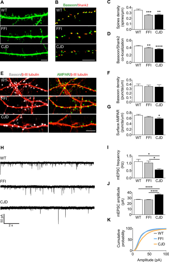
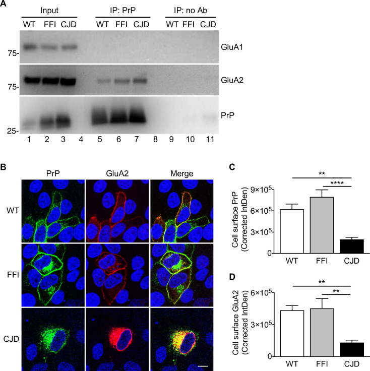
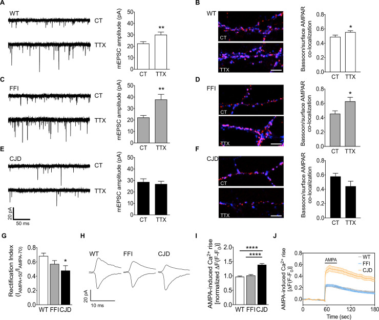
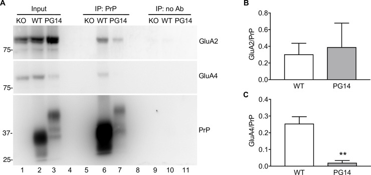
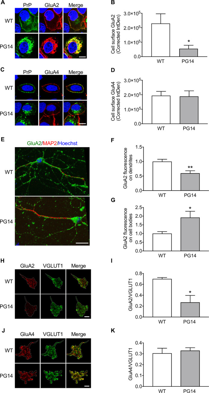
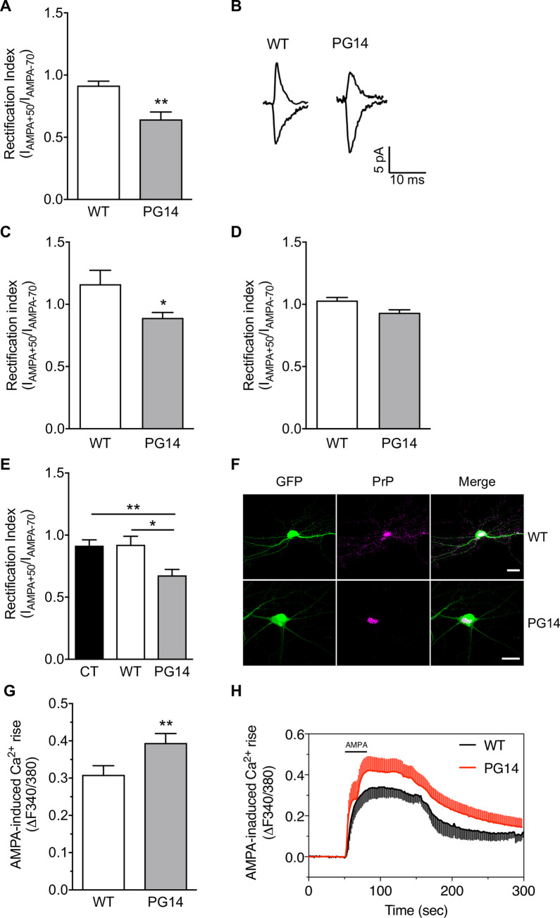
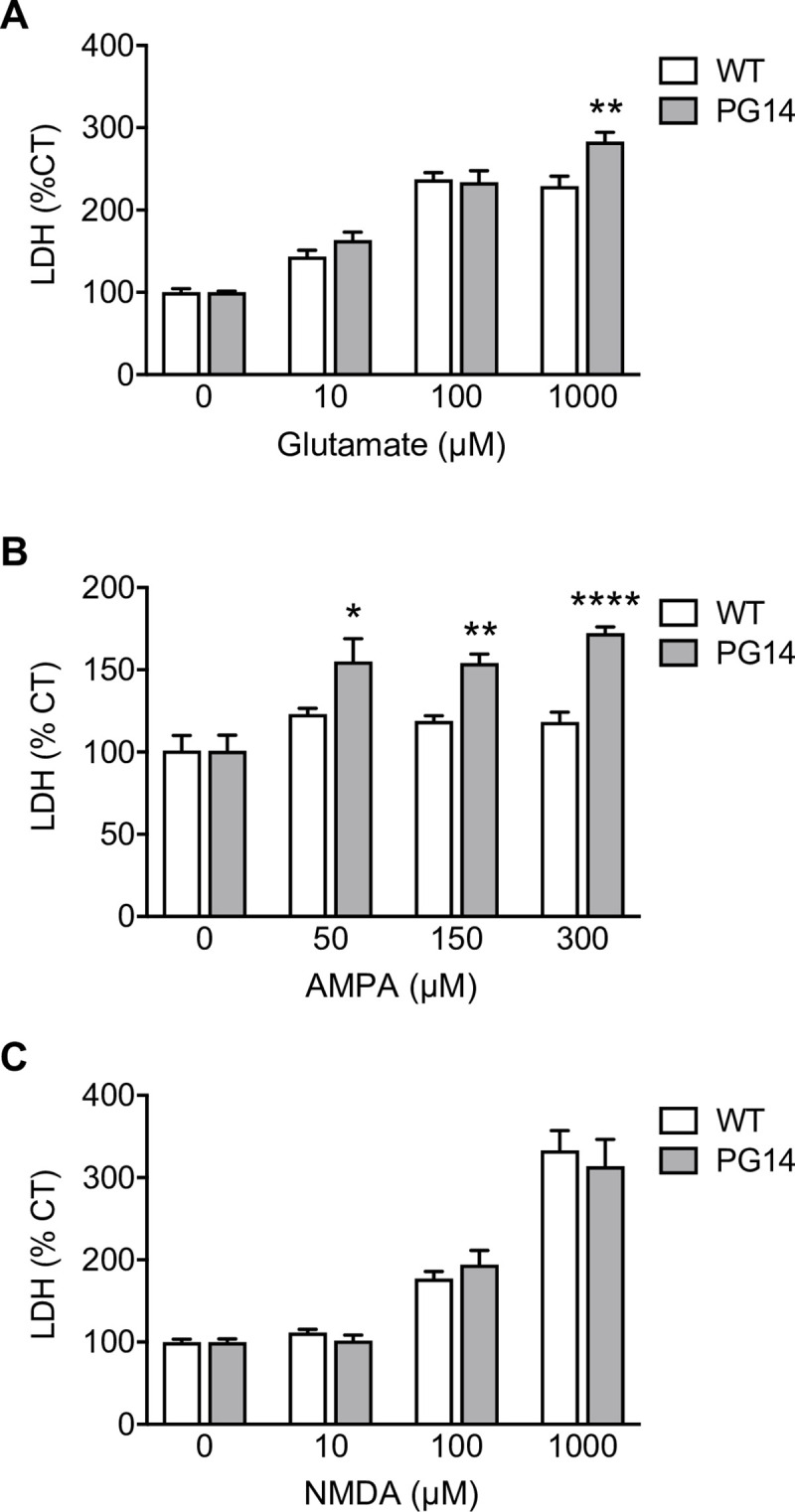
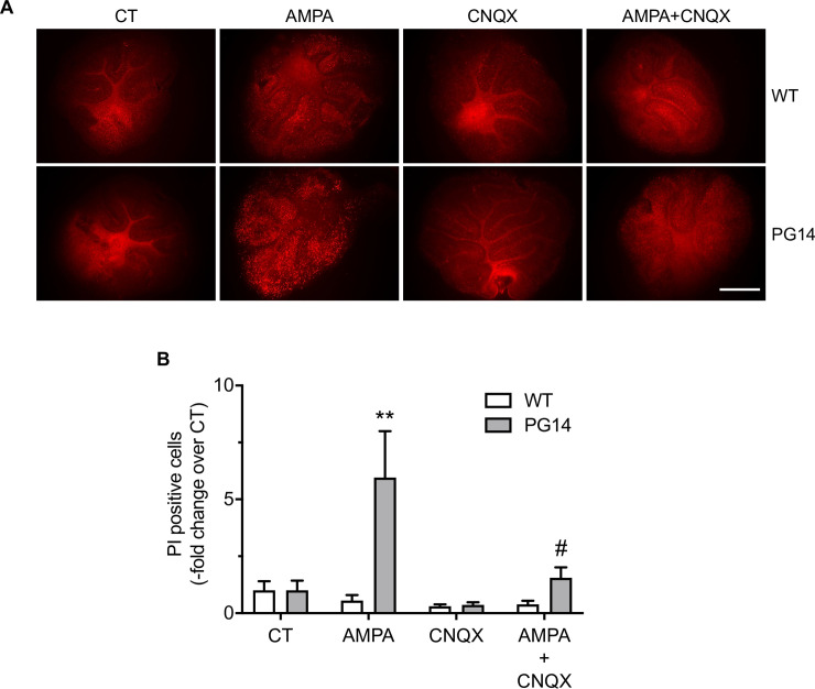
Prion protein (PrP) mutations are linked to genetic prion diseases, a class of phenotypically heterogeneous neurodegenerative disorders with invariably fatal outcome. How mutant PrP triggers neurodegeneration is not known. Synaptic dysfunction precedes neuronal loss but it is not clear whether, and through which mechanisms, disruption of synaptic activity ultimately leads to neuronal death. Here we show that mutant PrP impairs the secretory trafficking of AMPA receptors (AMPARs). Specifically, intracellular retention of the GluA2 subunit results in synaptic exposure of GluA2-lacking, calcium-permeable AMPARs, leading to increased calcium permeability and enhanced sensitivity to excitotoxic cell death. Mutant PrPs linked to different genetic prion diseases affect AMPAR trafficking and function in different ways. Our findings identify AMPARs as pathogenic targets in genetic prion diseases, and support the involvement of excitotoxicity in neurodegeneration. They also suggest a mechanistic explanation for how different mutant PrPs may cause distinct disease phenotypes.


Conflict of interest statement

The authors have declared that no competing interests exist.

Figures

Fig 1
Fig 1. Synaptic structure and activity are altered in FFI and CJD hippocampal neurons.
(A) Confocal representative images of WT, CJD and FFI neurons transfected with EGFP and (C) quantification of total spine density; 46–62 dendrites from 25–30 neurons for each condition. Kruskal-Wallis test followed by Dunn’s multiple comparison test: **p < 0.01, ***p < 0.001. (B) WT and mutant PrP neurons stained for Bassoon (green) and Shank2 (red) and (D) quantification of the levels of co-localization between the two; 69–87 fields for each condition, Kruskal-Wallis test followed by Dunn’s multiple comparison test: **p < 0.01, ****p < 0.0001. (E) WT, CJD and FFI neurons stained for Bassoon (gray), surface AMPAR (green) and β-III tubulin (red). Graphs show the number of Bassoon (F) or AMPAR (G) puncta per μm of dendrite; 17–23 dendrites from 10–15 neurons for each condition. One-way ANOVA followed by Tukey’s multiple comparison test: *p < 0.05. (H) Representative traces and analysis of mEPSC frequency (I) and cumulative amplitude (J, K). WT 32, FFI 25, CJD 18 cells. Kruskal-Wallis test followed by Dunn’s multiple comparison test: *p < 0.05, ****p < 0.0001. Data are the mean ± SEM of at least three independent experiments. Scale bars 5 μm in all images.
Fig 2
Fig 2. Mutant PrP interacts with the GluA2 AMPAR subunit impairing its membrane delivery.
(A) Hippocampal protein extracts (500 μg) from WT, FFI and CJD mice were incubated with uncoated magnetic beads (IP: no Ab) or magnetic beads coated with anti-PrP monoclonal antibody 94B4 (IP: PrP). The immunoprecipitated proteins were analyzed by western blot with anti-GluA1 or anti-GluA2 antibody or anti-PrP polyclonal antibody P45-66. The input is 20 μg of total proteins. Lanes 4 and 8 were left empty on purpose. This experiment is representative of three similar ones. (B) HeLa cells were co-transfected with plasmids encoding WT, FFI or CJD PrP-EGFP fusion protein, and the AMPAR subunit GluA2. After 48h, cells were fixed, permeabilized, stained with anti-GluA2 (red) antibody, and reacted with Hoechst 33258 (blue) to stain the nuclei. Scale bar 10 μm. (C, D) The fluorescent density of PrP and GluA2 on the cell surface was measured and background corrected (Corrected IntDen). Each bar indicates the mean ± SEM of 12–17 tranfected cells from three independent experiments. **p < 0.01, ****p < 0.0001 by one-way ANOVA, Tukey’s post hoc test.
Fig 3
Fig 3. AMPA receptor trafficking is impaired in CJD but not in FFI neurons.
(A, C, E) Representative traces and quantification of mEPSC amplitude in control and TTX-treated WT, FFI and CJD neurons. WT 16 (CT) and 18 (TTX), FFI 12 (CT) and 13 (TTX), CJD 18 (CT) and 16 (TTX) cells. Mann-Whitney test: **p < 0.01. (B, D, F) Confocal representative images and quantification of the levels of co-localization between Bassoon (blue) and surface AMPAR (red) in the conditions described above. Scale bar 5 μm. Unpaired Student’s t-test: *p < 0.05. (G) Rectification index (IAMPA+50/IAMPA-70) and (H) representative traces in WT and mutant neurons. WT 24, FFI 15, CJD 7 cells. Kruskal-Wallis test followed by Dunn’s multiple comparison test: *p < 0.05. (I) Analysis of calcium peaks and (J) representative traces of calcium response to AMPA (30 μM) in WT, FFI and CJD neurons. Six fields (79–150 responding cells) for each condition. Kruskal-Wallis test followed by Dunn’s multiple comparison test: ****p < 0.0001. Data are the mean ± SEM of at least three independent experiments.
Fig 4
Fig 4. PrP and GluA2 co-immunoprecipitate from cerebellar extracts.
(A) Cerebellar protein extracts (500 μg) from Tg(WT), Tg(PG14) and PrP knockout (KO) mice were incubated with uncoated magnetic beads (IP: no Ab) or magnetic beads coated with anti-PrP monoclonal antibody 94B4 (IP: PrP). The immunoprecipitated proteins were analyzed by western blot with anti-GluA2 or anti-GluA4 antibody or anti-PrP polyclonal antibody P45-66. The input is 20 μg of total proteins. (B, C) The amounts of immunoprecipitated GluA2 (B) and GluA4 (C) were quantified by densitometry of western blots like in (A) and normalized on the amount of immunoprecipitated PrP. Data are the mean ± SEM of 3–4 replicates from three independent experiments; **p < 0.01 by Student’s t-test.
Fig 5
Fig 5. GluA2 is retained intracellularly and is expressed less at post-synaptic sites in the granule cell layer of Tg(PG14) mice.
(A) HeLa cells were co-transfected with plasmids encoding WT or PG14 PrP-EGFP fusion protein, and the AMPAR subunit GluA2. After 48h cells were fixed, permeabilized, stained with anti-GluA2 (red), and reacted with Hoechst 33258 (blue) to stain the nuclei. (B) The fluorescent density of GluA2 on the cell surface was measured and corrected for background (Corrected IntDen). Each bar indicates the mean ± SEM of 12 WT and 14 PG14 PrP-transfected cells from three independent experiments; *p<0.01 by Student’s t-test. (C) HeLa cells were co-transfected with WT or PG14 PrP and EGFP-tagged GluA4. After 48 h cells were fixed, permeabilized, stained with anti-PrP monoclonal antibody 98A3 (green), and reacted with Hoechst 33258 (blue) to stain the nuclei. (D) The fluorescent density of GluA4 on the cell surface was measured and background corrected. Each bar indicates the mean ± SEM of 6 WT and 6 PG14 PrP-transfected cells. (E) CGNs from WT and PG14 mice were fixed, permeabilized and immunostained with mouse monoclonal anti-GluA2 (green) and chicken polyclonal anti-MAP2 (red). Cells were reacted with Hoechst 33258 (blue) to stain the nuclei, and analyzed by confocal microscopy using sequential Z-stack acquisition mode and 3D reconstruction. The GluA2 signal on dendrites (F) and cell bodies (G) was analyzed by NIH ImageJ software. Data are the mean ± SEM of 18 WT cells (28 dendrites) and 18 PG14 cells (27 dendrites) from three independent experiments. (H-K) Brain sections of WT and PG14 mice were stained with anti-GluA2 (H) or GluA4 (J) (red) and anti-VGLUT1 (green) antibodies. The GluA2 (I) and GluA4 (K) in each cerebellar glomerulus were measured and expressed as the ratio between the area of the AMPA receptor subunit and VGLUT1 signals. The dotted line identifies a single glomerulus used for quantification. Signal outside the ROI has been cleared with ImageJ for clarity. Bars indicate the mean ± SEM of three mice per group (1098–1392 glomeruli). *p < 0.05 by Student’s t-test. Scale bars 20 μm in A, C and E; and 125 μm in H and J.
Fig 6
Fig 6. PG14 PrP alters AMPA receptor subunit composition and calcium permeability in primary CGNs and transfected hippocampal cells.
(A) Rectification index (IAMPA+50/IAMPA-70) and (B) representative traces obtained by whole-cell patch-clamp experiments in WT and PG14 cultured cerebellar granule cells. I-70mV/I+50mV: WT 18, PG14 21. **p<0.01 by Mann-Whitney test. Rectification index recorded from cerebellar granule (C) or Purkinje (D) cells in acute brain slices. I-70mV/I+50mV granule cells: WT 8 cells from 3 mice; PG14 11 cells from 4 mice. I-70mV/I+50mV Purkinje cells: WT 4 cells from 3 mice; PG14 4 cells from 3 mice. * p<0.05 by Unpaired T-test. (E) Rectification index (IAMPA+50/IAMPA-70) in hippocampal neurons transfected with the pBud-CE4.1 vector expressing only GFP (CT), or also expressing WT or PG14 PrP. CT 18, WT 9, PG14 16 cells. Kruskal-Wallis test followed by Dunn’s multiple comparison test: *p < 0.05; **p < 0.01. (F) Confocal images showing primary hippocampal neurons from C57BL/6J mice transfected with a bigenic plasmid that drives efficient PrP and GFP expression. Scale bar 10 μm. (G) Analysis of calcium peaks and (H) representative traces of calcium response to AMPA (30 μM) in WT and PG14 cerebellar granule neurons. Six fields (102–123 responding cells) for each condition. **p < 0.01 by Mann-Whitney test. Bar graphs show mean ± SEM of at least of three independent experiments.
Fig 7
Fig 7. PG14 CGNs are hypersensitive to glutamate- and AMPA- but not NMDA-induced excitotoxicity.
CGNs from WT and PG14 mice were treated with glutamate (A), AMPA (B) or NMDA (C) at the concentrations indicated. After 24h cell death was quantified by LDH assay, and expressed as a percentage of the values for cells treated with the vehicle. Data are the mean ± SEM of 6–11 replicates from three independent experiments; *p < 0.05, **p < 0.01, ****p < 0.0001 by two-way ANOVA, Bonferroni’s post-hoc test.
Fig 8
Fig 8. Cultured organotypic cerebellar slices from Tg(PG14) mice show increased vulnerability to AMPA toxicity.
(A) Organotypic cerebellar slices from WT and PG14 mice were cultured for 15 days before exposure to 50 μM AMPA, 25 μM CNQX, or AMPA and CNQX, for 24h. Scale bar 500 μm. (B) The PI-positive cells were counted after 24h and expressed as the–fold change from vehicle-treated cells (CT). Data are the mean ± SEM of 9–24 cerebellar slices from three independent experiments. **p < 0.01 and vs. PG14 CT; #p < 0.05 vs. PG14 AMPA by two-way ANOVA, Tukey’s post-hoc test.

References

    1. Kim M-O, Takada LT, Wong K, Forner SA, Geschwind MD. Genetic PrP prion diseases. Cold Spring Harb Perspect Biol. 2018;10 10.1101/cshperspect.a033134 - DOI - PMC - PubMed
    1. Monari L, Chen SG, Brown P, Parchi P, Petersen RB, Mikol J, et al. Fatal familial insomnia and familial Creutzfeldt-Jakob disease: different prion proteins determined by a DNA polymorphism. Proc Natl Acad Sci U S A. 1994;91: 2839–42. 10.1073/pnas.91.7.2839 - DOI - PMC - PubMed
    1. Senatore A, Restelli E, Chiesa R. Synaptic dysfunction in prion diseases: a trafficking problem? Int J Cell Biol. 2013;2013: 543803 10.1155/2013/543803 - DOI - PMC - PubMed
    1. Steinert JR. Prion protein as a mediator of synaptic transmission. Commun Integr Biol. 2015;8: e1063753 10.1080/19420889.2015.1063753 - DOI - PMC - PubMed
    1. Senatore A, Colleoni S, Verderio C, Restelli E, Morini R, Condliffe SB, et al. Mutant PrP suppresses glutamatergic neurotransmission in cerebellar granule neurons by impairing membrane delivery of VGCC alpha(2)delta-1 subunit. Neuron. 2012;74: 300–13. 10.1016/j.neuron.2012.02.027 - DOI - PMC - PubMed

Publication types