. 2020 Sep;9(17):6354-6366.
doi: 10.1002/cam4.3303. Epub 2020 Jul 16.

Exosome-transmitted linc00852 associated with receptor tyrosine kinase AXL dysregulates the proliferation and invasion of osteosarcoma

Affiliations

Exosome-transmitted linc00852 associated with receptor tyrosine kinase AXL dysregulates the proliferation and invasion of osteosarcoma

Qiming Li et al. Cancer Med. 2020 Sep.

Abstract

Background: Receptor tyrosine kinase AXL has been found to be highly expressed in osteosarcoma and positively associated with poor prognosis. There are tumor groups with high or low AXL expression, which had different capabilities of invading vessels and forming distal metastases. Exosome-transmitted lncRNA may be transferred intercellularly to promote tumor cells' proliferation and invasion.

Methods: Exosomes were detected by electron microscopy, particle size analysis, and western blotting. High-throughput sequencing helped to find the highest differentially expressed lncRNA in AXL-associated exosomes. Clone formation, wound healing, transwell assay, and xenograft model in nude mice were performed to evaluate cells' proliferation, migration, and invasion in vitro and in vivo. Lentiviral transfection was used to up- or down-regulate the lncRNA levels in cell lines. Luciferase reporter assay and RNA FISH etchelped to indicate the molecular mechanisms. The results in the cell lines were proved in the osteosarcoma tissues with clinical analysis.

Results: The exosomes derived from donor cells with high AXL expression could promote the proliferation and invasion and upregulate AXL expression of the receiver cells with low AXL. Linc00852 was the highest differentially expressed lncRNA in AXL-associated exosomes and was also regulated by AXL expression. Although the mechanisms of linc00852 in nucleus were unrevealed, it could upregulate AXL expression partly by competitively binding to miR-7-5p. The AXL-exosome-linc00852-AXL positive feedback loop might exist between the donor cells and the receiver cells. Clinically, linc00852 was significantly highly expressed in osteosarcoma tissues and positively associated with tumor volumes and metastases, which was also obviously related with AXL mRNA expression.

Conclusion: AXL-associated exosomal linc00852 up-regulated the proliferation, migration, and invasion of osteosarcoma cells, which would be considered as a new tumor biomarker and a special therapeutic target for osteosarcoma.

Keywords: AXL; exosome; invasion; linc00852; osteosarcoma.


Conflict of interest statement

It has been submitted with the full knowledge and approval of the First Affiliated Hospital, Sun Yat‐sen University. All authors have significantly contributed and are in agreement with the content of the final manuscript. The authors declare that there are no conflict of interest in connection with the work submitted.

Figures

FIGURE 1
FIGURE 1
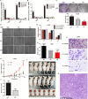
Characterization of exosomes derived from osteosarcoma cells with high AXL expression and the functions of the exosomes after being transmitted to receiver cells with low AXL expression. A, Electron microscopy images of exosomes derived from osteosarcoma cells 143B and HOS with high and low AXL expression. B, The size distribution of exosomes derived from 143B and HOS were detected by a particle size analysis; the particle size ranged from 40 to 200 nm, accounting for 51.3% and 67.1%, respectively. C, Exosomes derived from 143B and HOS cells were analyzed by flow cytometry with CD63 and CD81. D, Exosomes were analyzed by Western blot with TSG101 and Alix. E, Wound healing assays showed that the migration ability of 143B cells with low AXL expression increased significantly after co‐culturing with the exosomes derived from cells with high AXL expression for 24 h. F, Similarly, the exosomes also increased the migration ability of HOS. G,H, The Transwell assays showed the migration abilities of 143B and HOS. The number of migrated cells with high AXL expression was significantly higher than the number of migrated cells with low AXL expression. Meanwhile, the number of the migrated cells with low AXL expression increased significantly after 24 h of co‐culturing with the exosomes derived from the cells with high AXL expression. I, The Matrigel‐coated Boyden chamber assays showed the invasive ability of 143B and HOS cells with low AXL expression similarly increased significantly with treatment of the exosomes. J, The growth curves of 143B with different AXL levels in nude mice. The xenograft tumors with high AXL expression grew faster than the tumors with low AXL expression. After treating with the exosomes derived from the cells with high AXL expression, the growth rate of the xenograft tumors with low AXL expression increased significantly. K, The weight of xenograft tumors with high AXL expression was greater than that of the tumors with low AXL expression. The weight of xenograft tumors of cells co‐cultured with the exosomes increased significantly the weight of tumors with low AXL expression. L, H&E staining showed that the pathological features of xenograft tumors were similar to the ordinary osteosarcoma. One lung metastatic foci occurred in the group of cells co‐cultured with the exosomes compared with no metastasis of the untreated xenograft tumors. (*P < .05, **P < .01, ***P < .001)
FIGURE 2
FIGURE 2
Exosomes derived from donor cells with high AXL expression promoted the expression of AXL mRNA and protein in receiver cells with low AXL expression, and linc00852 was the highest differentially expressed lncRNA in AXL‐associated exosomes. A, AXL mRNA expression by qRT‐PCR detection was significantly higher in cells with high AXL expression than in cells with low AXL expression by flow cytometry for both 143B and HOS. B, The AXL mRNA expression of groups with low AXL expression increased significantly both for 143B and HOS after cells with low AXL expression were co‐cultured with exosomes derived from cells with high AXL expression 24 h. C, AXL protein expression was similar to mRNA by Western blot. D, High‐throughput sequencing analysis revealed that there were many lncRNAs increased in the exosomes from 143B with highly expressed AXL (right) compared with the exosomes from 143B with AXL low expression (left), among which linc00852 was the highest differentially expressed lncRNA. E, The linc00852 expression by qRT‐PCR detection in osteosarcoma cell lines was significantly higher than that in NIH3T3 cells, and linc00852 was detected at relatively high levels in 143B and HOS. F, The expression of linc00852 in the cells with high AXL expression was significantly higher than in the cells with low AXL expression. G, The expression of linc00852 in exosomes derived from cells with high AXL expression was significantly higher than in cells with low AXL expression. H, The expression of linc00852 in the exosomes decreased dramatically after being treated with 2 μg/ml RNase and Triton 0.1%X‐100, whereas there was no obvious change with the treatment of RNase alone in both 143B and HOS cells. I, The linc00852 expression of the cells with low AXL expression increased significantly both for 143B and HOS after they were co‐cultured with exosomes derived from cells with high AXL expression for 24 h. (*P < .05, **P < .01, ***P < .001)
FIGURE 3
FIGURE 3
Exosomal linc00852 promoted proliferation, migration and invasion of osteosarcoma cells in vitro and in vivo. A, The transfection efficiency was detected by qRT‐PCR. Linc00852 was highly expressed in both 143B and HOS cells transfected with lentiviral vectors encoding the full linc00852 sequence. Data are shown as the mean ± SD. B, AXL mRNA expression of transfected 143B and HOS cells were detected by qRT‐PCR. The trends of AXL expression were similar to the linc00852 expression. C, Clone formation assays showed that in 143B the clone numbers were increased significantly in over‐linc00852 cells and decreased significantly in SH‐linc00852 cells compared with the control. D, The migration ability of 143B cells was detected with wound healing assays. E, Compared with the control, the migration distance of over‐linc00852 cells significantly decreased, and the migration distance of SH‐linc00852 cells significantly increased after 24 h. F, The invasive potential of 143B cells was detected by Matrigel‐coated Transwell assays. G, Compared with the control, the invasive ability of over‐linc00852 cells was significantly enhanced. H, The growth curve of transfected 143B cells in nude mice. Over‐linc00852 xenograft tumors grew significantly faster than the NC‐linc00852 group. I, The size of the xenograft tumors in the over‐linc00852 group was significantly larger than in the control group. J, The weight of the xenograft tumors in the over‐linc00852 group was significantly greater than in the control group. K, H&E staining showed that the pathological features of the xenograft tumors were similar to ordinary osteosarcoma. (*P < .05, **P < .01, ***P < .001)
FIGURE 4
FIGURE 4
Over‐linc00852 increased the expression of AXL and AKT protein, and linc00852 upregulated AXL partly by competing for miR‐7‐5p binding. A, The Western blot analysis showed that the AXL and AKT protein levels increased in over‐linc00852 143B cells. B, The target scan analysis revealed the binding site of linc00852 and miR‐7‐5p and the primer sequence. C, According to qRT‐PCR the expression of miR‐7‐5p in cells with high AXL expression was significantly lower than that in cells with low AXL expression. D, The WT‐linc00852 vector (or Mut‐linc00852) and miR‐7‐5p mimics were co‐transfected into 293T cells; the miR‐7‐5p mimic‐transfected cells reduced the luciferase activity of WT‐linc00852 vector compared with the mutant vector. E, The same results of linc00852 and miR‐7‐5p were further confirmed in 143B cells. F, The WT‐AXL vector (or Mut‐AXL) and miR‐7‐5p mimics were co‐transfected into 293T cells. The miR‐7‐5p mimic reduced the luciferase activities of the AXL WT reporter vector but not the AXL mutant reporter vector. G, The same results of AXL and miR‐7‐5p were further confirmed in 143B cells. (*P < .05, **P < .01, ***P < .001)
FIGURE 5
FIGURE 5
linc00852 located mainly in the nucleus of osteosarcoma cells and the possible mechanism of AXL‐exosome‐linc00852‐AXL positive feedback loop. A, RNA FISH of 143B and HOS cells showed that linc00852 was primarily located in the nucleus, and only a small number was located in the cytoplasm.U6 was used as the nuclear control. B, The qRT‐PCR of cellular fractions further confirmed that linc00852showedintracellular localization. C, Schematic diagram of exosomal linc00852‐mediated osteosarcoma growth and progression. Donor cells with high AXL expression secreted exosomal linc00852; then, the exosomes were transmitted to receiver cells with low AXL expression. A few linc00852 might function as a ceRNA for miR‐7‐5p to facilitate AXL expression in the cytoplasm while most linc00852 might transfer to the receiver cells’ nucleus and increase the expression of AXL with unknown complex mechanisms, sequentially activating the AKT pathway to promote the progression of the receiver cells.
FIGURE 6
FIGURE 6
The expression of linc00852, AXL mRNA and miR‐7‐5p in osteosarcoma tissues and the relationship among them. A, The expression of linc00852 in osteosarcoma tissues and adjacent tissues in patients. B, linc00852 levels were significantly higher in osteosarcoma tissues than in the adjacent normal tissues from 34 patients. C, linc00852 expression in tumors with a diameter greater than 8 cm increased significantly versus in the tumors smaller than 8 cm. D, linc00852 expression in tumors with metastasis increased significantly compared with the tumors without metastasis. E, The Kaplan‐Meier analysis revealed that patients with higher linc00852 expression had lower disease‐free survival (DFS). F, Patients with higher linc00852 also had lower overall survival (OS). G, The expression of miR‐7‐5p in osteosarcoma tissues and adjacent tissues in patients. H, miR‐7‐5p levels were significantly lower in osteosarcoma tissues than in the adjacent normal tissues. I, AXL levels were significantly higher in osteosarcoma tissues than in the adjacent normal tissues. J, In the clinical specimens, there was a positive relationship between AXL mRNA and linc00852. K, There was a negative correlation trend with no significance between miR‐7‐5p and linc00852 in osteosarcoma. L, There was a negative correlation trend with no significance between AXL mRNA and miR‐7‐5p in osteosarcoma

Similar articles

Cited by

References

    1. Jiang N, Wang X, Xie X, et al. lncRNA DANCR promotes tumor progression and cancer stemness features in osteosarcoma by upregulating AXL via miR‐33a‐5p inhibition. Cancer Lett. 2017;405:46‐55. - PubMed
    1. Simpson S, Dunning MD, de Brot S, Grau‐Roma L, Mongan NP, Rutland CS. Comparative review of human and canine osteosarcoma: morphology, epidemiology, prognosis, treatment and genetics. Acta Vet Scand. 2017;59(1):71. - PMC - PubMed
    1. Rankin EB, Giaccia AJ. The receptor tyrosine kinase AXL in cancer progression. Cancers. 2016;8(11):103. - PMC - PubMed
    1. Zhu C, Wei Y, Wei X. AXL receptor tyrosine kinase as a promising anti‐cancer approach: functions, molecular mechanisms and clinical applications. Mol Cancer. 2019;18(1):153. - PMC - PubMed
    1. Antony J, Huang RY. AXL‐driven EMT state as a targetable conduit in cancer. Cancer Res. 2017;77(14):3725‐3732. - PubMed

Publication types

MeSH terms