Review
. 2020 Jul;10(7):200118.
doi: 10.1098/rsob.200118. Epub 2020 Jul 15.

Modulators of calcium signalling at fertilization

Affiliations
Review

Modulators of calcium signalling at fertilization

Paula Stein et al. Open Biol. 2020 Jul.

Abstract

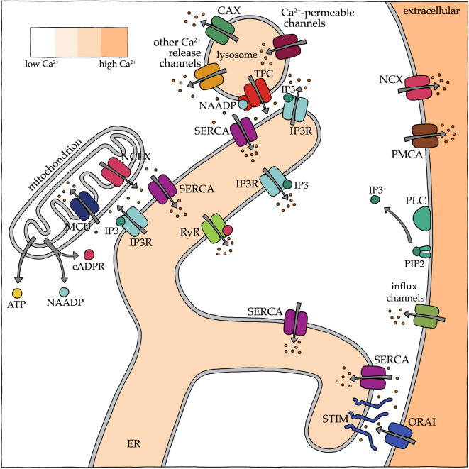
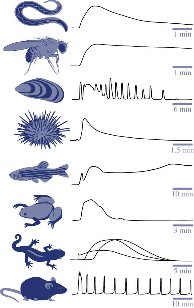
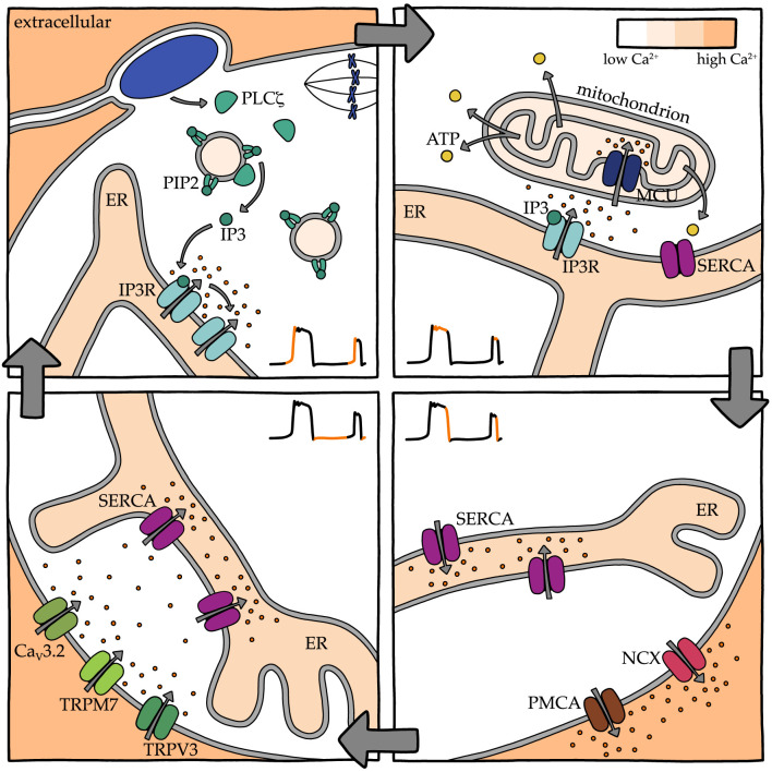
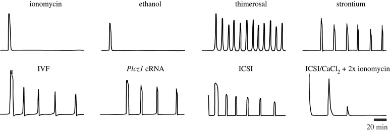
Calcium (Ca2+) signals initiate egg activation across the animal kingdom and in at least some plants. These signals are crucial for the success of development and, in the case of mammals, health of the offspring. The mechanisms associated with fertilization that trigger these signals and the molecules that regulate their characteristic patterns vary widely. With few exceptions, a major contributor to fertilization-induced elevation in cytoplasmic Ca2+ is release from endoplasmic reticulum stores through the IP3 receptor. In some cases, Ca2+ influx from the extracellular space and/or release from alternative intracellular stores contribute to the rise in cytoplasmic Ca2+. Following the Ca2+ rise, the reuptake of Ca2+ into intracellular stores or efflux of Ca2+ out of the egg drive the return of cytoplasmic Ca2+ back to baseline levels. The molecular mediators of these Ca2+ fluxes in different organisms include Ca2+ release channels, uptake channels, exchangers and pumps. The functions of these mediators are regulated by their particular activating mechanisms but also by alterations in their expression and spatial organization. We discuss here the molecular basis for modulation of Ca2+ signalling at fertilization, highlighting differences across several animal phyla, and we mention key areas where questions remain.

Keywords: calcium channels; calcium signalling; egg activation; fertilization; oocyte.


Conflict of interest statement

We declare we have no competing interests.

Figures

Figure 1.
Figure 1.
Elements of Ca2+ toolbox in endoplasmic reticulum, mitochondria, lysosomes and plasma membrane of somatic cells. Orange dots indicate Ca2+; grey arrows show direction of Ca2+ flow. cADPR, cyclic ADP ribose; CAX, Ca2+/proton exchanger; IP3, inositol trisphosphate; IP3R, IP3 receptor; MCU, mitochondrial uniporter; NCLX, mitochondrial sodium–Ca2+ exchanger; NAADP, nicotinic acid adenine dinucleotide phosphate; NCX, sodium/Ca2+ exchanger; ORAI, Ca2+ release-activated Ca2+ channel protein; PIP2, phosphatidylinositol 4,5-bisphosphate; PLC, phospholipase C; PMCA, plasma membrane Ca2+ ATPase; RyR, ryanodine receptor; SERCA, sarco/endoplasmic reticulum Ca2+ ATPase pump; STIM, stromal interaction molecule; TPC, two-pore channel.
Figure 2.
Figure 2.
Diagrams of typical cytoplasmic Ca2+ changes at fertilization across various species. From top to bottom: nematode (Caenorhabditis elegans) [62], fruit fly (Drosophila melanogaster) [63], purple mussel (Septifer virgatus) [7], sea urchin (Paracentrotus lividus) [64], zebrafish (Danio rerio) [65], frog (Xenopus laevis) [66], newt (Cynops pyrrhogaster) [67] and mouse (Mus musculus) [68]. Time indicated on scale bar for each Ca2+ trace. The three lines on the newt trace represent spatially distinct signals induced by different sperm during physiological polyspermy. Traces adapted from indicated references.
Figure 3.
Figure 3.
Generation of a Ca2+ wave. Localized production of IP3 (teal circles) by PLC (teal semicircles) leads to IP3-mediated Ca2+ release from clusters of IP3 receptors in endoplasmic reticulum (ER) membranes. The released Ca2+ promotes Ca2+-induced Ca2+ release from nearby clusters, increasing the cytoplasmic Ca2+ gradient. In addition, the released Ca2+ stimulates PLC to generate additional IP3 in a positive feedback loop. Continued IP3 production and Ca2+ diffusion lead eventually to Ca2+ release from distal IP3 receptor clusters in distinct ER regions and a wave of Ca2+ release across the cell. Orange dots indicate Ca2+; grey arrows show the direction of IP3 or Ca2+ flow.
Figure 4.
Figure 4.
Cycle of Ca2+ transient generation in mammalian eggs at fertilization. Starting at the top left, the large grey arrows show temporal order. For each panel, the cytoplasmic Ca2+ trace is coloured orange at the portions of the trace that are generated mainly due to the steps illustrated in that panel. Top left: Sperm PLCζ acts on PIP2 in intracellular vesicles to generate IP3, which stimulates IP3R-mediated Ca2+ release and subsequent Ca2+-induced Ca2+ release. Top right: Ca2+ stimulates mitochondrial ATP production; ATP is required for SERCA pump activity. Bottom right: Ca2+ is pumped back into the ER through SERCA pumps and out of the egg through PMCA pumps and NCX. Bottom left: Ca2+ flows into the cytoplasm through TRMP7, CaV3.2 and TRPV3 channels and is then available for SERCA pumps to replenish ER Ca2+ stores in preparation for the next Ca2+ release event. Orange dots indicate Ca2+ at its destination; small grey arrows show the direction of flow. CaV3.2, T-type voltage-dependent Ca2+ channel; IP3, inositol trisphosphate; IP3R, IP3 receptor; MCU, mitochondrial uniporter; NCX, sodium/Ca2+ exchanger; PIP2, phosphatidylinositol 4,5-bisphosphate; PLCζ, phospholipase C zeta; PMCA, plasma membrane Ca2+ ATPase; SERCA, sarco/endoplasmic reticulum Ca2+ ATPase pump; TRPM7, transient receptor potential cation channel subfamily M member 7; TRPV3, transient receptor potential cation channel subfamily V member 3.
Figure 5.
Figure 5.
Representative Ca2+ traces resulting from egg activation by various methods. Top row, chemical inducers of artificial egg activation (AEA) in bovine eggs [234]. Bottom row, in vitro fertilization (IVF) [68], Plcz1 cRNA injection [123] and intracytoplasmic sperm injection (ICSI) [235] in mouse eggs; ICSI using sperm lacking PLCζ activity, in medium containing CaCl2, followed by two treatments with ionomycin in human egg [235]. Scale bar applies to all traces. Traces adapted from indicated references.

References

    1. Lillie F. 1919. Problems of fertilization. Chicago, IL: University of Chicago Press.
    1. Dalcq A. 1928. Le rôle du calcium et du potassium dans l'entrée en maturation de l'oeuf de pholade (Barnea candida). Protoplasma 4, 18–44. (10.1007/BF01607955) - DOI
    1. Ridgway EB, Gilkey JC, Jaffe LF. 1977. Free calcium increases explosively in activating medaka eggs. Proc. Natl Acad. Sci. USA 74, 623–627. (10.1073/pnas.74.2.623) - DOI - PMC - PubMed
    1. Gilkey JC, Jaffe LF, Ridgway EB, Reynolds GT. 1978. A free calcium wave traverses the activating egg of the medaka, Oryzias latipes. J. Cell Biol. 76, 448–466. (10.1083/jcb.76.2.448) - DOI - PMC - PubMed
    1. Shimomura O, Johnson FH, Saiga Y. 1962. Extraction, purification and properties of aequorin, a bioluminescent protein from the luminous hydromedusan, Aequorea. J. Cell. Comp. Physiol. 59, 223–239. (10.1002/jcp.1030590302) - DOI - PubMed

Publication types

MeSH terms

Substances

LinkOut - more resources