Review
. 2020 Jul 17;127(3):427-447.
doi: 10.1161/CIRCRESAHA.120.316958. Epub 2020 Jul 16.

Myofibroblasts and Fibrosis: Mitochondrial and Metabolic Control of Cellular Differentiation

Affiliations
Review

Myofibroblasts and Fibrosis: Mitochondrial and Metabolic Control of Cellular Differentiation

Andrew A Gibb et al. Circ Res. .

Abstract

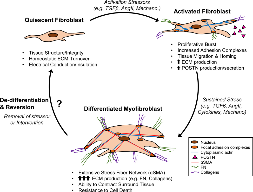
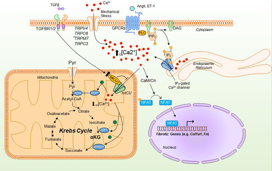
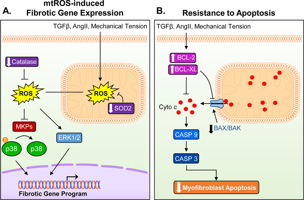
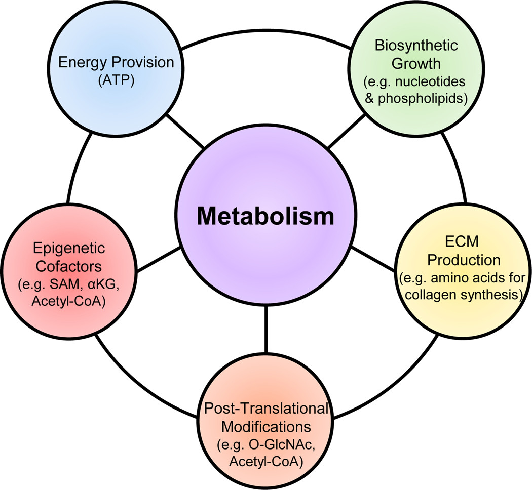
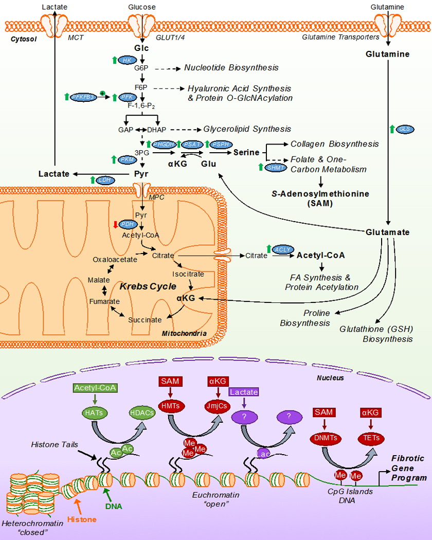
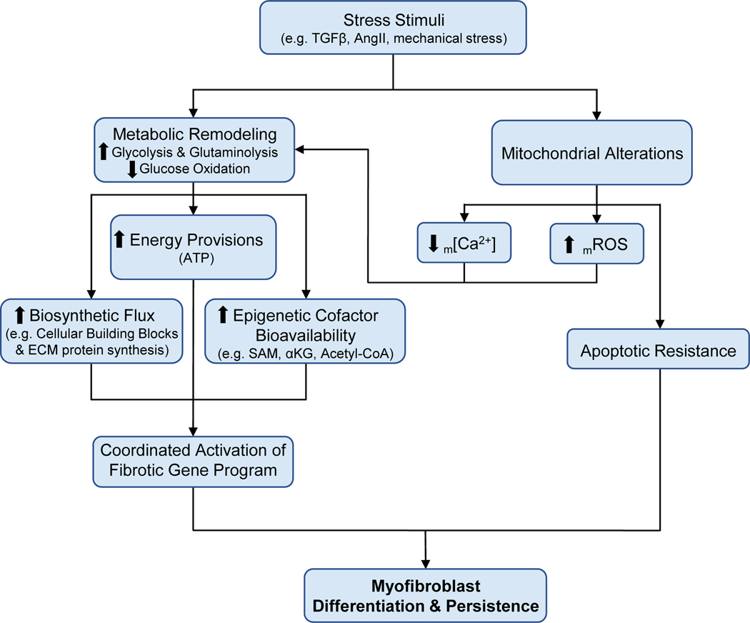
Cardiac fibrosis is mediated by the activation of resident cardiac fibroblasts, which differentiate into myofibroblasts in response to injury or stress. Although myofibroblast formation is a physiological response to acute injury, such as myocardial infarction, myofibroblast persistence, as occurs in heart failure, contributes to maladaptive remodeling and progressive functional decline. Although traditional pathways of activation, such as TGFβ (transforming growth factor β) and AngII (angiotensin II), have been well characterized, less understood are the alterations in mitochondrial function and cellular metabolism that are necessary to initiate and sustain myofibroblast formation and function. In this review, we highlight recent reports detailing the mitochondrial and metabolic mechanisms that contribute to myofibroblast differentiation, persistence, and function with the hope of identifying novel therapeutic targets to treat, and potentially reverse, tissue organ fibrosis.

Keywords: fibrosis; heart failure; metabolism; mitochondria; myofibroblast.


Figures

Figure 1.
Figure 1.. Progression and characterization of cardiac fibroblast to myofibroblast conversion.
Resident fibroblasts contribute to cardiac homeostasis under normal physiological conditions. Upon increased mechanical tension and pro-fibrotic mediators (e.g. TGFβ, AngII), these resident cardiac fibroblasts become activated, at which time they infiltrate and expand at the site of injury as well as begin to remodel the ECM. Upon sustained and unremitting activation signaling, these fibroblasts differentiate into myofibroblasts characterized by the de novo expression of αSMA and the excessive production of ECM proteins (FN, collagens). Myofibroblasts are also resistant to cell death, resulting in their persistence in the injured heart, eventually leading to maladaptive tissue remodeling. Recent evidence suggest that myofibroblasts are capable of de-differentiating upon removal of stress stimuli, however the mechanisms by which they do so, and the potential for targeted mechanistic interventions, require further investigation. ECM – extracellular matrix; TGFβ – transforming growth factor beta; AngII – angiotensin II; Mechano – mechanical stress; POSTN – periostin; αSMA – α-smooth muscle actin; FN – fibronectin.
Figure 2.
Figure 2.. Mechanistic actions of Ca2+ during myofibroblast differentiation.
Mechanical activation of TRP channels permits Ca2+ entry from the extracellular space, increasing cytosolic [Ca2+]. Additionally, activation of the Gq/PLC/IP3 pathway allows for IP3-mediated Ca2+ release from the ER, further increasing cytosolic [Ca2+]. This increase in cytosolic [Ca2+] activates calmodulin-dependent calcineurin phosphatase (CaM/CN) to dephosphorylate and permit nuclear translocation of NFAT and subsequent activation of the fibrotic gene program. While mitochondria can act to buffer increases in cytosolic [Ca2+] through the mitochondrial calcium uniporter complex (mtCU), the transcriptional upregulation of MICU1 alters mtCU gating, reducing mCa2+ uptake and reducing m[Ca2+]. As m[Ca2+] is an activator of numerous dehydrogenases and phosphatases within the mitochondria, this significantly reduces pyruvate oxidation and overall Krebs cycle flux. TGFβ – transforming growth factor beta; AngII – angiotensin II; ET-1 – endothelin 1; TGFBR1/2 – TGFβ receptor 1/2; TRPs – transient receptor potential channels; PLC – protein lipase C; PIP2 – phosphatidylinositol 4,5-bisphosphate; DAG – diacylglycerol; Pyr – pyruvate; PDH – pyruvate dehydrogenase; PDP – pyruvate dehydrogenase phosphatase; MICU1 – mitochondrial calcium uptake 1; NFAT – nuclear factor of activated T-cells; IDH – isocitrate dehydrogenase; ⍺KGDH - ⍺-ketoglutarate dehydrogenase; Col1a1 – collagen 1 type 1; Fn – fibronectin.
Figure 3.
Figure 3.. Mitochondrial mechanisms of myofibroblast differentiation and persistence.
(A) Pro-fibrotic stressors increase ROS production in the mitochondria, which then results in an increased and sustained intracellular ROS load, in part, through the downregulation of SOD2 and catalase. Increases in intracellular ROS in turn activates p38 and ERK1/2 signaling pathways, which are known to increase the transcription of the fibrotic gene program. (B) A key feature of myofibroblasts is their resistance to apoptosis. Mitochondrial cytochrome c (Cyto c) release is prevented through the upregulation of anti-apoptotic factors (BLC-2, BCL-XL) while pro-apoptotic factors (BAX, BAK) are downregulated. Reduced activation of the proteolytic caspase cascade due to decreased cytochrome c release from the mitochondria contributes to the persistence of myofibroblasts in the injured heart by imparting a resistance to cell death. TGFβ – transforming growth factor beta; AngII – angiotensin II; ROS – reactive oxygen species; SOD2 - superoxide dismutase 2; MKPs – mitogen-activated protein kinases; BCL-2 – B-cell lymphoma 2; BCL-XL - B-cell lymphoma-extra large; BAX – Bcl-2-associated X protein; BAK – Bcl-2 homologous antagonist killer; Casp9/3 – caspase 9/3.
Figure 4.
Figure 4.. Metabolic components of cellular differentiation.
Overview of the metabolic influence regulating fibroblast differentiation. In addition to the energetic demands of a proliferating/differentiating cell, it is necessary to increase the biosynthetic intermediates to support such events. An increase in production and secretion of ECM components (e.g. collagens) requires increased amino acid synthesis. Post-translational modifications of proteins (e.g. transcription factors) can greatly alter their activities while regulation of the epigenetic landscape influences gene program activation & silencing. While each can work independently, the integration of these metabolic actions drive and coordinate cellular differentiation. ECM – extracellular matrix; SAM – S-adenosylmethionine; ⍺KG - ⍺-ketoglutarate.
Figure 5.
Figure 5.. Metabolic inputs regulating the biosynthetic and transcriptional changes coordinating myofibroblast differentiation.
General schematic of myofibroblast metabolism on the differentiation program. Myofibroblasts are characterized by an increase in glutaminolysis and aerobic glycolysis, accompanied by decreased glucose oxidation. These key metabolic re-programming events permit the use of carbon intermediates by ancillary biosynthetic pathways to coordinate the production of ECM constituents, protein modifications, and epigenetic cofactors that initiate and sustain the myofibroblast differentiation program. Changes in the expression/activity of noted enzymes along with metabolites in bold are key to the differentiation process. Of these processes, the ability of these key metabolites to act as cofactors for epigenetic modifying enzymes is critical for the coordinated activation and silencing of transcriptional events which promote the fibrotic gene program and myofibroblast differentiation/persistence. αKG – α-ketoglutarate; Pyr – pyruvate; HK – hexokinase; PFK – phosphofructokinase; PFKFB3 – 6-phosphofructo-2-kinase/fructose-2,6-bisphosphatase 3; PHGDH – phosphoglycerate dehydrogenase; PSAT – phosphoserine amino transferase; PSPH – phosphoserine phosphatase; PKM – pyruvate kinase muscle isozyme; LDH – lactate dehydrogenase; SHMT – serine hydroxymethyltransferase; PDH – pyruvate dehydrogenase; ACLY – ATP citrate lyase; HATs – histone acetyltransferases; HDACs – histone deacetylases; HMT – histone methyltransferases; JmJCs – Jumonji C-domain-containing histone demethylases; DNMTs – DNA methyltransferases; TETs – ten-eleven translocases; Ac – acetylation mark; Me – methylation mark; Lac – lactylation mark.
Figure 6.
Figure 6.. Working model of the influence of mitochondria and metabolism on myofibroblast differentiation and persistence.
Pro-fibrotic stress stimuli are critical for the activation and differentiation of resident cardiac fibroblasts to myofibroblasts. Pro-fibrotic stimulation results in significant metabolic remodeling that is critical for the energy provisions and synthesis of cellular building blocks required for proliferation, growth, and differentiation. Furthermore, these changes in cellular metabolism also regulate the bioavailability of metabolites which serve as cofactors for numerous epigenetic-modifying enzymes to coordinate the activation of the fibrotic gene program to promote differentiation. Recent work from our lab has shed new light on the mitochondria as a key regulator to this process by reducing mitochondrial Ca2+ uptake to promote the metabolic remodeling required for the differentiation process. The mitochondria also increase the generation of mROS to promote the necessary metabolic remodeling. Additionally, mitochondria under pro-fibrotic stimulation and in the terminally differentiated state downregulate the apoptotic pathways, promoting myofibroblast persistence which contributes to the progressive nature of cardiovascular disease. Collectively, the coordinated efforts of mitochondrial and metabolic remodeling are critical for the activation, differentiation, and persistence of myofibroblasts in cardiac disease, providing novel therapeutic targets to mitigate and potential reverse tissue fibrosis in disease. TGFβ – transforming growth factor beta; AngII – angiotensin II; m[Ca2+] - mitochondrial calcium concentration; mROS – mitochondrial-derived reactive oxygen species; ECM – extracellular matrix; SAM – S-adenosylmethionine; ⍺KG - ⍺-ketoglutarate.

References

    1. Diez J. Mechanisms of cardiac fibrosis in hypertension. J Clin Hypertens (Greenwich). 2007;9:5. - PMC - PubMed
    1. Humeres C and Frangogiannis NG. Fibroblasts in the Infarcted, Remodeling, and Failing Heart. JACC Basic Transl Sci. 2019;4:449–467. - PMC - PubMed
    1. Bonnans C, Chou J and Werb Z. Remodelling the extracellular matrix in development and disease. Nat Rev Mol Cell Biol. 2014;15:786–801. - PMC - PubMed
    1. Michel G, Tonon T, Scornet D, Cock JM and Kloareg B. The cell wall polysaccharide metabolism of the brown alga Ectocarpus siliculosus. Insights into the evolution of extracellular matrix polysaccharides in Eukaryotes. New Phytol. 2010;188:82–97. - PubMed
    1. Theocharis AD, Skandalis SS, Gialeli C and Karamanos NK. Extracellular matrix structure. Adv Drug Deliv Rev. 2016;97:4–27. - PubMed

Publication types