Review
. 2020 Aug;38(8):835-845.
doi: 10.1016/j.tibtech.2020.01.006. Epub 2020 Mar 5.

Engineering Heart Morphogenesis

Affiliations
Review

Engineering Heart Morphogenesis

Christian J Mandrycky et al. Trends Biotechnol. 2020 Aug.

Abstract

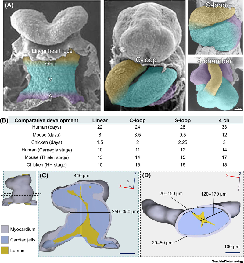
Recent advances in stem cell biology and tissue engineering have laid the groundwork for building complex tissues in a dish. We propose that these technologies are ready for a new challenge: recapitulating cardiac morphogenesis in vitro. In development, the heart transforms from a simple linear tube to a four-chambered organ through a complex process called looping. Here, we re-examine heart tube looping through the lens of an engineer and argue that the linear heart tube is an advantageous starting point for tissue engineering. We summarize the structures, signaling pathways, and stresses in the looping heart, and evaluate approaches that could be used to build a linear heart tube and guide it through the process of looping.

Keywords: biomaterials; heart tube looping; mechanobiology; organogenesis; stem cells; tissue engineering.


Figures

Figure 1.
Figure 1.. Blueprints of the lopping human heart tube.
(A) SEM images of the developing human heart tube from linear hear tube, C-looped, S-looped, and four-chambered heart (4 ch, bottom right). Future outflow tracts (OFT; yellow), ventricles (V; blue), and atria (A; purple) are color coded to highlight the complex structural transition the heart tube undergoes during morphogenesis. Color coding is a rough anatomical estimation and not exact. Images of the linear and C-loop heart tube stages were adapted from UNSW Embryology (https://embryology.med.unsw.edu.au), and S-loop and 4 chamber stages were adapted from [75]. (B) Comparison of the heart’s developmental timeline in different vertebrates by days post-fertilization and speciesspecific developmental stages for human (Carnegie), mouse (Thieler [76]), and chick (Hamburger-Hamilton (HH) [2]). Comparisons were based on whole embryo developmental stages and do not always reflect the relative stages of cardiac development for each species. (C) 3D reconstruction of the human linear heart tube at stage 10 viewed with a Z-axis or longitudinal cross-section. Measurements of the tube’s height and diameter from the outer-most myocardial walls are overlaid. (D) Y-axis or crosswise section of the linear heart tube and thickness ranges of the myocardial wall (grey), lumen (yellow), and cardiac jelly (blue) layers. In each reconstruction, the surrounding coelomic walls, foregut, or neural tubes are excluded for clarity. All reconstructions and overlaid measurements in (C) and (D) are adapted with permission from the 3D Atlas of Human Embryology (specimen 6330) [77]. Multiple (n=5) measurements of each structure were taken along the z-axis from each cross-sectional view plane in (C) and (D) to create a range of size values. Measurements were taken from a 3D-rendered PDFs using the snap-to-edge tool which is not as accurate as using more precise 3D rendering software but are provided to give an approximate range.
Figure 2.
Figure 2.. Spatiotemporal patterns of gene expression for heart looping may be engineered in vitro.
(A) Select signaling pathways and factors involved in the development of the looping heart tube that can potentially be controlled using existing techniques. The looping heart tube can be broadly divided into four areas based on what part of the heart they will form: outflow tract, future ventricles, future atria, and future atrioventricular canal. Cells in each of those areas have unique gene and protein expression profiles that allow them to specialize in performing their specific tasks. While previously published reviews described these expression profiles in detail [,–,– 41], here we have identified the few that can potentially be controlled using currently existing techniques (specific reference numbers provided in figure image). (B) Techniques for controlling heart tube looping-associated pathways. We have identified three main existing approaches to modulating signaling pathways in engineered or explanted native tissues: controlled diffusion chambers for creating gradients of soluble factors, functionalized hydrogels, and optogenetic methods. Diffusion-based techniques rely on controlling incubation chamber geometry to prevent convective flows, controlling medium flow in a microfluidic chamber, or both. Functional hydrogels allow for photopatterning and photorelease of a variety of factors with micron-level resolution. Additionally, they allow for control of the mechanical properties of local cell environment via photomediated gel cross-linking or degradation. Finally, optogenetic methods rely on genetic modifications of the cells allowing for spatiotemporal up- and downregulation of specific expression factors in those cells using light.
Figure 3.
Figure 3.. Mechanobiological cues are critical in heart tube morphogenesis.
(A) Forces acting on the heart tube can arise from (1) boundary conditions and reaction forces at the inflow at outflow tracts, (2) cellular forces from the growth, proliferation, and contraction of the cells of the heart tube, (3) hemodynamic forces from the flow of blood through the heart tube, and (4) forces from the growth and extension of the embryo and organs that border the heart tube. (B) The result of these loads on the heart tube are deformations that contribute to C-looping, including extension and bending at the heart-foregut interface, bending due to cell differential cell growth and contraction, and distention of the heart tube lumen due to hemodynamic pressure changes. Color in (B) represents strain given the applied tissue deformation.

References

    1. Wallingford JB (2019) The 200-year effort to see the embryo. Science 365, 758–759 - PubMed
    1. Hamburger V and Hamilton HL (1992) A series of normal stages in the development of the chick embryo. 1951. Dev. Dyn. Off. Publ. Am. Assoc. Anat 195, 231–272 - PubMed
    1. Becker JR et al. (2011) Human cardiomyopathy mutations induce myocyte hyperplasia and activate hypertrophic pathways during cardiogenesis in zebrafish. Dis. Model. Mech 4, 400–410 - PMC - PubMed
    1. Borchardt T and Braun T (2007) Cardiovascular regeneration in non-mammalian model systems: what are the differences between newts and man? Thromb. Haemost 98, 311–318 - PubMed
    1. Ivanovitch K et al. (2017) Live imaging of heart tube development in mouse reveals alternating phases of cardiac differentiation and morphogenesis. eLife 6, e30668. - PMC - PubMed

Publication types