Review
. 2020 Aug;38(8):857-872.
doi: 10.1016/j.tibtech.2020.01.003. Epub 2020 Feb 18.

Engineering Microphysiological Immune System Responses on Chips

Affiliations
Review

Engineering Microphysiological Immune System Responses on Chips

Chris P Miller et al. Trends Biotechnol. 2020 Aug.

Abstract

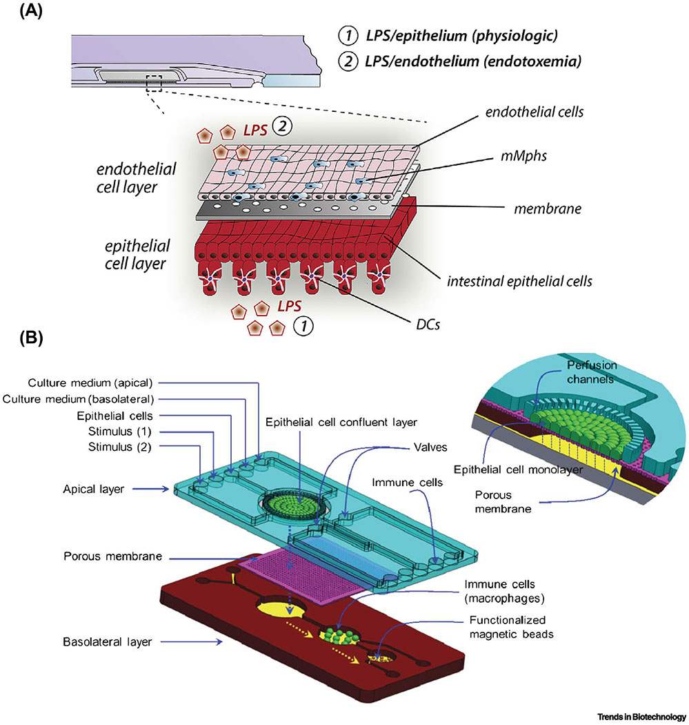
Tissues- and organs-on-chips are microphysiological systems (MPSs) that model the architectural and functional complexity of human tissues and organs that is lacking in conventional cell monolayer cultures. While substantial progress has been made in a variety of tissues and organs, chips recapitulating immune responses have not advanced as rapidly. This review discusses recent progress in MPSs for the investigation of immune responses. To illustrate recent developments, we focus on two cases in point: immunocompetent tumor microenvironment-on-a-chip devices that incorporate stromal and immune cell components and pathomimetic modeling of human mucosal immunity and inflammatory crosstalk. More broadly, we discuss the development of systems immunology-on-a-chip devices that integrate microfluidic engineering approaches with high-throughput omics measurements and emerging immunological applications of MPSs.

Keywords: engineering immune system responses; immune system-on-a-chip; intestinal inflammation; microphysiological systems; systems immunology; tumor immune microenvironment.


Conflict of interest statement

Declaration of Interests

D.H.K. is a scientific founder and equity holder of NanoSurface Biomedical Inc. H.J.K. is a founder of 3D Health Solutions Inc. and holds an equity interest in the company.

Figures

Figure 1.
Figure 1.. MPS modeling of the infiltration and cytotoxicity of therapeutic lymphocytes.
(A) To study the influence of monocytes on tumor cell killing by TCR-engineered T cells in the tumor microenvironment, a simple and customizable microfluidic device was developed for the 3D culture of HepG2 hepatocellular carcinoma tumor spheroids expressing the preS1 portion of the hepatitis B virus envelope protein linked to green fluorescent protein (preS1-GFP) and monocytes in a central collagen hydrogel. This was flanked by two media channels with or without TCR-engineered T cells specific for the hepatitis B virus antigen present on the tumor cells. (B) Tumor cell killing was assessed by confocal imaging of pre-labeled tumor spheroids (green) surrounded by monocytes (blue) and T cells (white). Dead tumor target cells were labeled by the live/dead cell exclusion dye DRAQ7 (red), and the volume of dead cells was quantified relative to the total spheroid volume. Reproduced from [16] under a Creative Commons license.
Figure 2.
Figure 2.. MPS modeling of the ADCC of NK cells in the tumor microenvironment.
(A) Tumor spheroids were embedded in a collagen hydrogel containing endothelial-lined lateral lumens and antibodies and NK cells were administered via the lumens or by direct inclusion in the collagen. (B) The inset shows how an immunocytokine combining an epithelial-directed antibody (anti-EpCAM) with a co-stimulating cytokine (IL-2) via IL-2 receptor (IL-2 R) enhances NK-cell cytotoxicity. (C) The steps in fabrication and cell seeding of the microfluidic device. (D) Photograph of actual microarray device labeled with a blue dye for visualization. (E) Confocal (top), the orthogonal cross-sectional view (bottom) and 3D reconstruction (right) of an endothelial lumen fixed and stained with an antibody for the endothelial marker CD31 (green), Hoechst (nucleus, blue), and phalloidin (actin, red). (F) The microfluidic device allowed the simultaneous detection of prelabeled human breast cancer MCF7 cells in a spheroid (blue), the epithelial marker EpCAM on the surface (red), and NK cells (green). (G) A hypoxia-sensing dye (red) confirmed increased hypoxia in the central region of a GFP-labeled MCF7 cell spheroid. Reproduced from [24] with permission.
Figure 3.
Figure 3.. MPS modeling of complex cell types in the tumor immune microenvironment.
(A) In one approach, patient tumor organoids can be harvested to retain the autologous stromal cells and immune cells prior to embedding in 3D microfluidic hydrogel devices. Confocal imaging of a patient high-grade serous ovarian carcinoma organoid containing viable (calcein acetoxymethyl ester (AM), green), EpCAM+ epithelial cells (purple) demonstrated the presence of CD8+ T cells (red) and all nucleated cells (Hoechst, blue). Microfluidic devices have facilitated the identification of cytokine profiles predictive of differential sensitivity to PD-1 blockade and exogenous agents that enhance sensitivity. Adapted from [28] under a Creative Commons license. (B) To provide for tunable control over cell density, type, ratio, and orientation, a “tumor ecosystem-on-a-chip” was developed (top). Confocal imaging of one half of the device demonstrated the position of pre-labeled human BT474 breast cancer cells (green), PBMC including immune cells (blue), and human breast Hs578T CAF (red) parallel to a central HUVEC monolayer (magenta). Magnified 3D images show physical interactions between various cell types (bottom). (C) Experimental design of the microfluidic devices containing two hydrogel compartments (gray) containing the indicated cell type configurations to allow for the study of how ADCC stimulated by the therapeutic antibody trastuzumab (Trast) is antagonized by CAF. Adapted from [30] under a Creative Commons license.
Figure 4.
Figure 4.. MPS modeling of intestinal inflammation.
(A) A schematic of the human gut inflammation-on-a-chip that provides the accessibility and versatility to manipulate the complex inflammatory milieu. Arrows in the central microchannels and the vacuum chambers indicate the directions of fluid flow and the peristalsis-like mechanical distortions, respectively. AP, apical; BL, basolateral. (B) Top-down views of the fluorescently labeled villous epithelium under the oxidative stress in the presence or absence of dextran sodium sulfate at various combinations of inflammatory triggers (PBMC, LPS, and E. coli) in the human gut inflammation-on-a-chip. Bar, 50 μm. (C) Cross-sectional views of the immune cell recruitment visualized in situ in the gut inflammation-on-a-chip. Gray, microengineered villous epithelium; green, PBMC. Dotted lines indicate the contour of the 3D villi. Dashed lines show the location of the basement membrane in the gut inflammation-on-a-chip. Bars, 50 μm. B and C reproduced from [60] with permission.
Figure 5.
Figure 5.. Immunocompetent intestinal MPS models that incorporate differentiated immune subsets.
(A) An MPS model that induces inflammatory responses in the EMI axis consisting of the intestinal epithelium, endothelium, LPS or bacterial cells, and tissue-resident mucosal macrophages (mMphs) and dendritic cells (DCs). Reproduced from [59] with permission. (B) A NutriChip that assesses the effects of dairy products by reconstituting intestinal epithelium, LPS or dairy food products, and macrophages. Bars, 50 μm. Reproduced from [61] with permission.

References

    1. Arrowsmith J and Miller P (2013) Trial watch: phase II and phase III attrition rates 2011-2012. Nat Rev Drug Discov 12 (8), 569. - PubMed
    1. Tsui JH et al. (2018) Conductive silk-polypyrrole composite scaffolds with bioinspired nanotopographic cues for cardiac tissue engineering. J Mater Chem B 6 (44), 7185–7196. - PMC - PubMed
    1. Smith AST et al. (2019) NanoMEA: A tool for high-throughput, electrophysiological phenotyping of patterned excitable cells. Nano Letters doi: 10.1021/acs.nanolett.9b04152. [Epub ahead of print]. - DOI - PMC - PubMed
    1. Nam KH et al. (2016) Multiscale cues drive collective cell migration. Sci Rep 6, 29749. - PMC - PubMed
    1. Park J et al. (2019) Switch-like enhancement of epithelial-mesenchymal transition by YAP through feedback regulation of WT1 and Rho-family GTPases. Nat Commun 10 (1), 2797. - PMC - PubMed

Publication types