. 2020 Oct;159(4):1357-1374.e10.
doi: 10.1053/j.gastro.2020.06.088. Epub 2020 Jul 13.

Activating Transcription Factor 6 Mediates Inflammatory Signals in Intestinal Epithelial Cells Upon Endoplasmic Reticulum Stress

Affiliations

Activating Transcription Factor 6 Mediates Inflammatory Signals in Intestinal Epithelial Cells Upon Endoplasmic Reticulum Stress

Stephanie T Stengel et al. Gastroenterology. 2020 Oct.

Abstract

Background & aims: Excess and unresolved endoplasmic reticulum (ER) stress in intestinal epithelial cells (IECs) promotes intestinal inflammation. Activating transcription factor 6 (ATF6) is one of the signaling mediators of ER stress. We studied the pathways that regulate ATF6 and its role for inflammation in IECs.

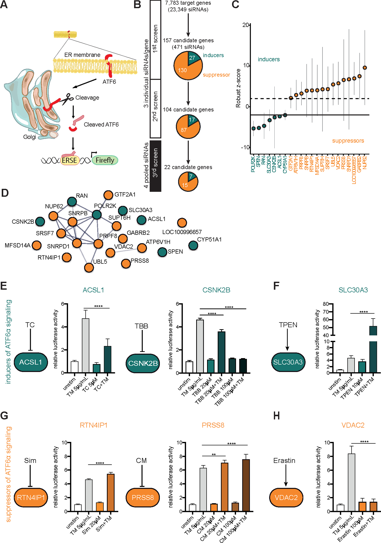
Methods: We performed an RNA interference screen, using 23,349 unique small interfering RNAs targeting 7783 genes and a luciferase reporter controlled by an ATF6-dependent ERSE (ER stress-response element) promoter, to identify proteins that activate or inhibit the ATF6 signaling pathway in HEK293 cells. To validate the screening results, intestinal epithelial cell lines (Caco-2 cells) were transfected with small interfering RNAs or with a plasmid overexpressing a constitutively active form of ATF6. Caco-2 cells with a CRISPR-mediated disruption of autophagy related 16 like 1 gene (ATG16L1) were used to study the effect of ATF6 on ER stress in autophagy-deficient cells. We also studied intestinal organoids derived from mice that overexpress constitutively active ATF6, from mice with deletion of the autophagy related 16 like 1 or X-Box binding protein 1 gene in IECs (Atg16l1ΔIEC or Xbp1ΔIEC, which both develop spontaneous ileitis), from patients with Crohn's disease (CD) and healthy individuals (controls). Cells and organoids were incubated with tunicamycin to induce ER stress and/or chemical inhibitors of newly identified activator proteins of ATF6 signaling, and analyzed by real-time polymerase chain reaction and immunoblots. Atg16l1ΔIEC and control (Atg16l1fl/fl) mice were given intraperitoneal injections of tunicamycin and were treated with chemical inhibitors of ATF6 activating proteins.

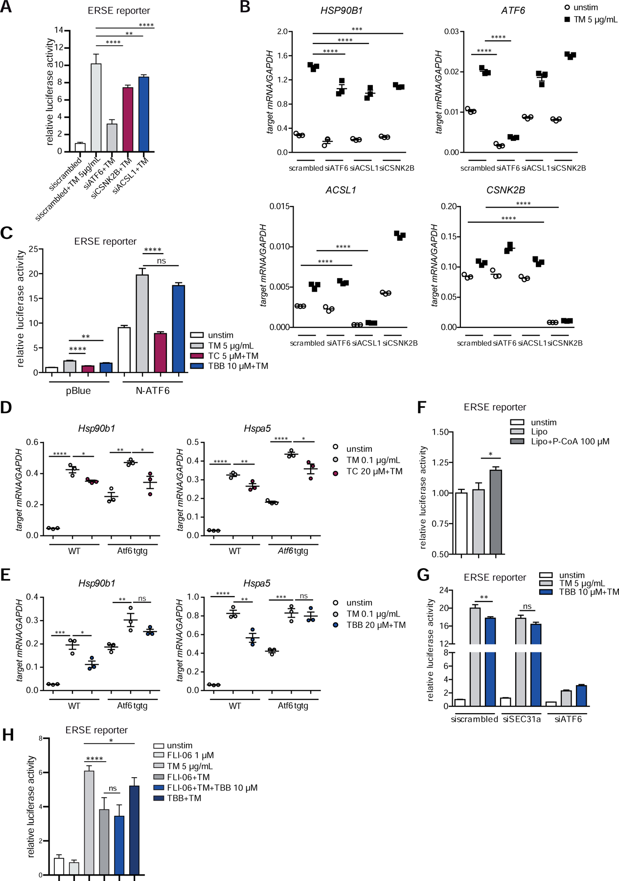
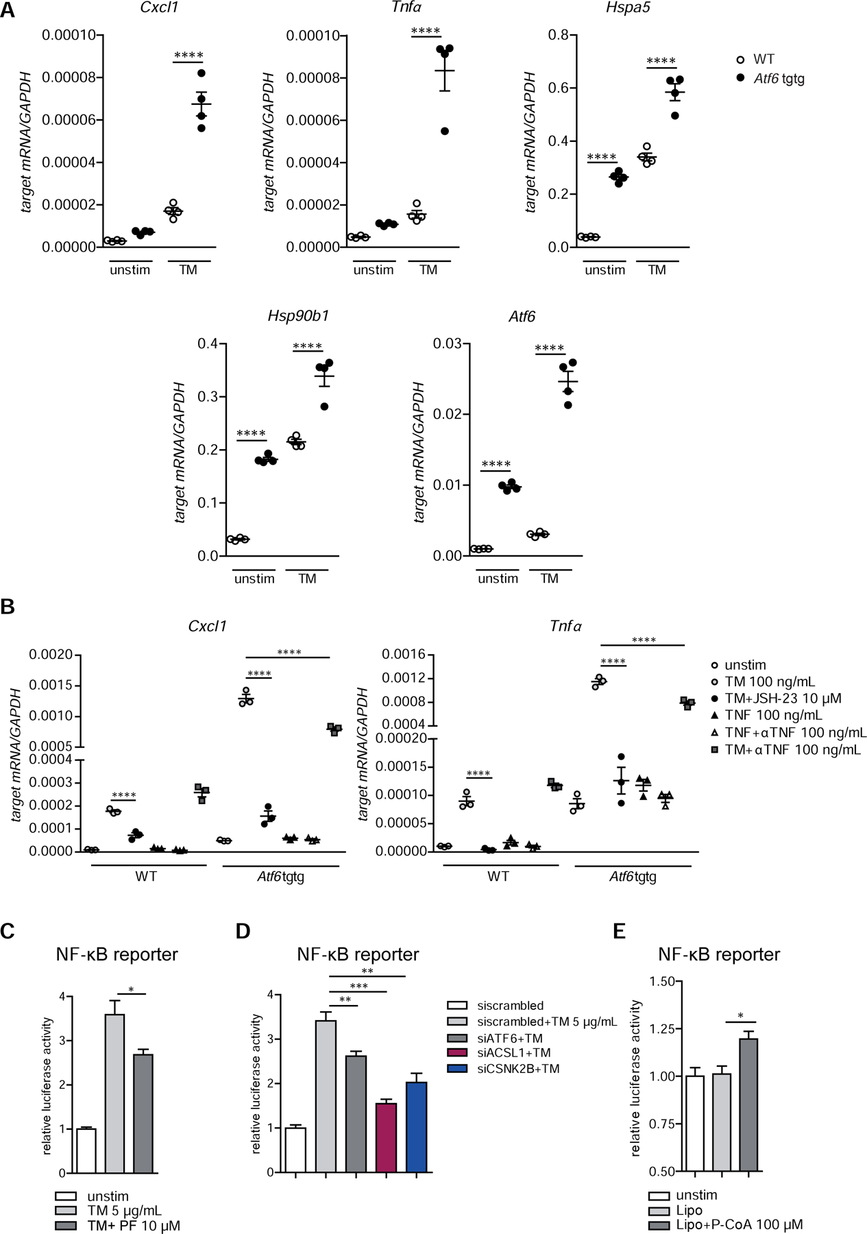
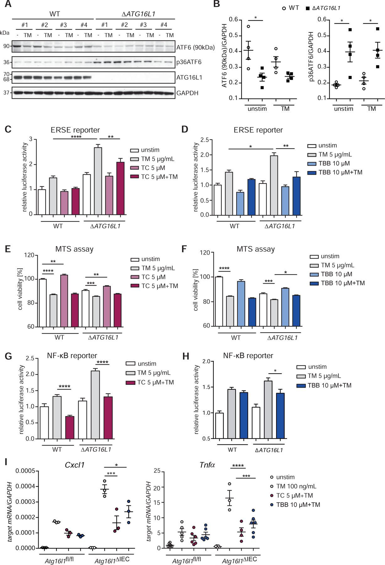
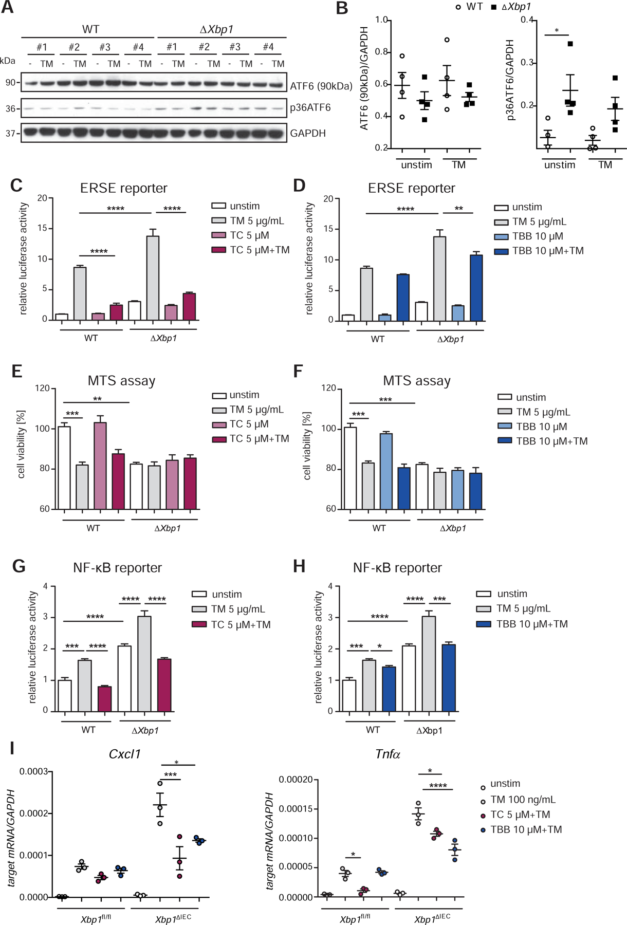
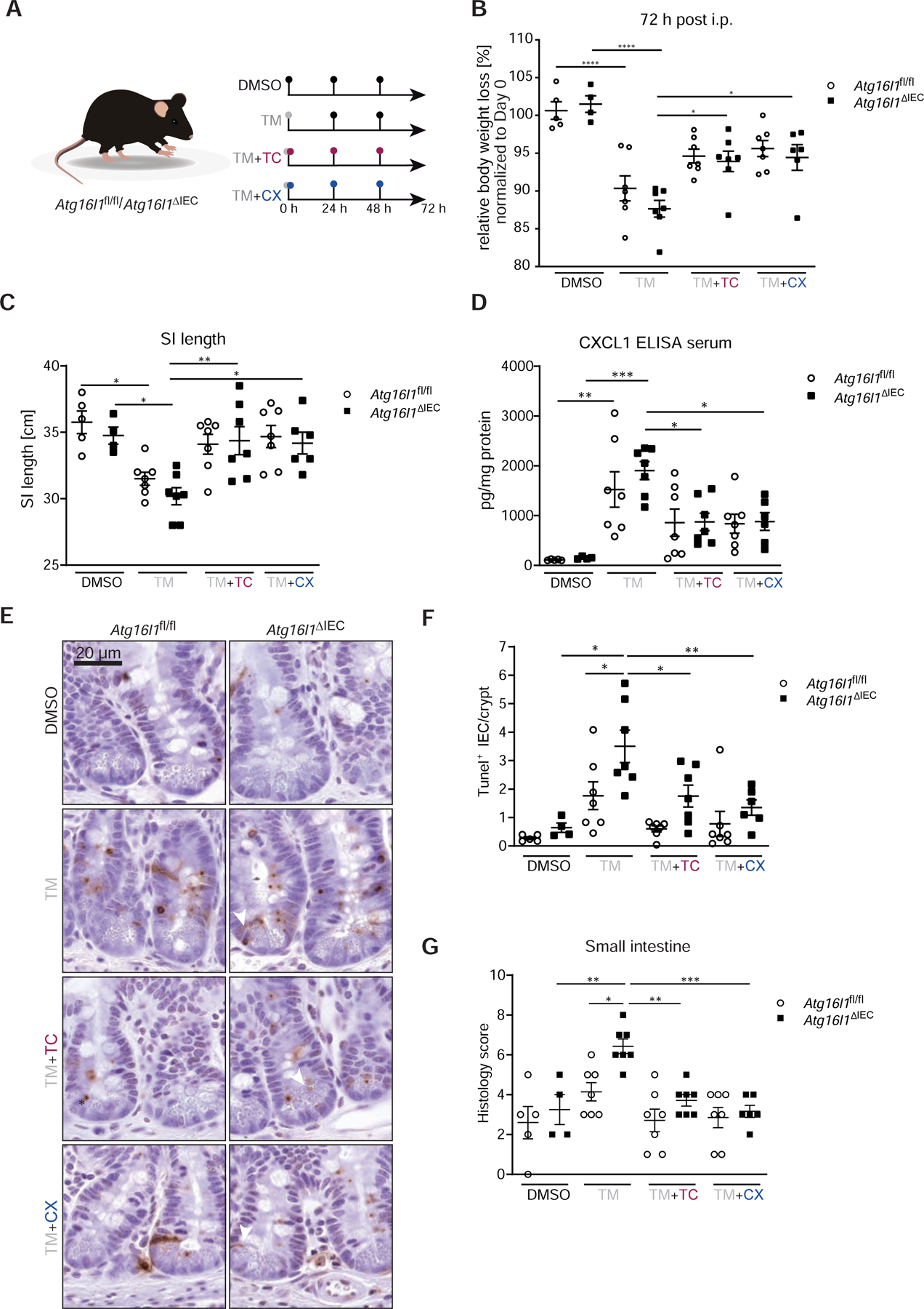
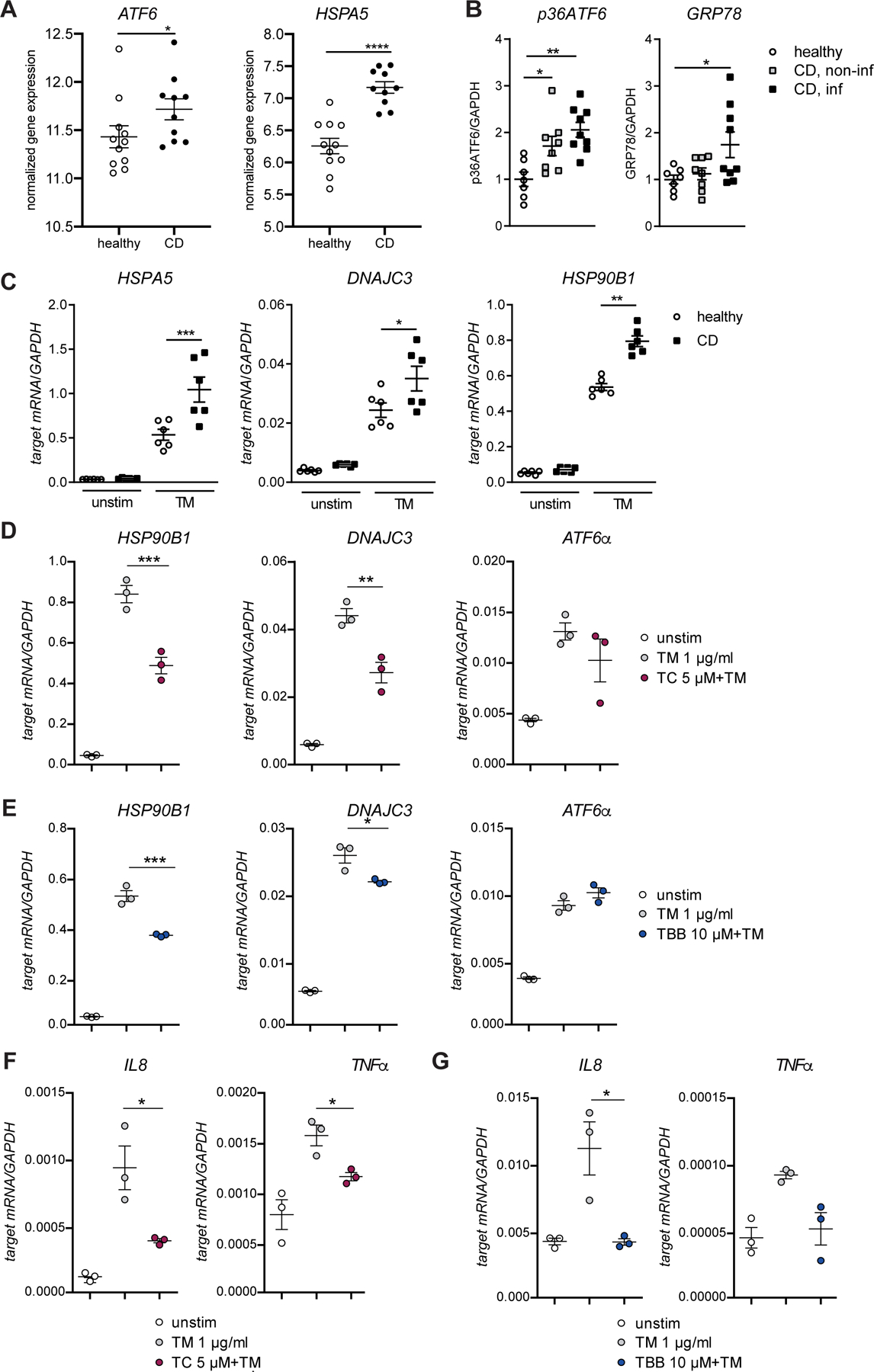
Results: We identified and validated 15 suppressors and 7 activators of the ATF6 signaling pathway; activators included the regulatory subunit of casein kinase 2 (CSNK2B) and acyl-CoA synthetase long chain family member 1 (ACSL1). Knockdown or chemical inhibition of CSNK2B and ACSL1 in Caco-2 cells reduced activity of the ATF6-dependent ERSE reporter gene, diminished transcription of the ATF6 target genes HSP90B1 and HSPA5 and reduced NF-κB reporter gene activation on tunicamycin stimulation. Atg16l1ΔIEC and or Xbp1ΔIEC organoids showed increased expression of ATF6 and its target genes. Inhibitors of ACSL1 or CSNK2B prevented activation of ATF6 and reduced CXCL1 and tumor necrosis factor (TNF) expression in these organoids on induction of ER stress with tunicamycin. Injection of mice with inhibitors of ACSL1 or CSNK2B significantly reduced tunicamycin-mediated intestinal inflammation and IEC death and expression of CXCL1 and TNF in Atg16l1ΔIEC mice. Purified ileal IECs from patients with CD had higher levels of ATF6, CSNK2B, and HSPA5 messenger RNAs than controls; early-passage organoids from patients with active CD show increased levels of activated ATF6 protein, incubation of these organoids with inhibitors of ACSL1 or CSNK2B reduced transcription of ATF6 target genes, including TNF.

Conclusions: Ileal IECs from patients with CD have higher levels of activated ATF6, which is regulated by CSNK2B and HSPA5. ATF6 increases expression of TNF and other inflammatory cytokines in response to ER stress in these cells and in organoids from Atg16l1ΔIEC and Xbp1ΔIEC mice. Strategies to inhibit the ATF6 signaling pathway might be developed for treatment of inflammatory bowel diseases.

Keywords: Gene Expression; IBD; Inflammation; Signal Transduction.


Conflict of interest statement

Conflict of interest: The authors disclose no conflicts.

Figures

Figure 1:
Figure 1:. Systematic siRNA screening reveals modulators of ATF6α activation.
(A) Schematic representation of the screening approach. (B) Screening procedure and number of candidates at different screening stages. In the primary and secondary screens each gene was targeted with three individual siRNAs tested separately. Pools of four siRNAs were used for the third screen. Candidate genes with an inducing or repressing effect on ATF6 activation are indicated in green or orange, respectively (C) Final set of 22 candidates after the third screen. Bars depict mean and 95th confidence interval (3 replicates). (D) STRING Network of candidate genes (third screen). Only interactions with a confidence score >0.4 were considered. Inducers depicted in green, repressors shown in orange. (EH) ERSE promoter activity quantified by dual luciferase reporter assay in HEK-293 cells. n=6. Cells were exposed to tunicamycin (5 µg/ml) and small molecule agents for 24 h. Depicted data representative of 3 independent experiments. Statistical analysis was performed using one-way ANOVA together with Tukey post hoc test. TC=Triacsin C; TBB=4,5,6,7-Tetrabromo-2-azabenzimidazole; TPEN=N,N,N′,N′-Tetrakis(2-pyridylmethyl)ethylenediamine; CM=Camostat mesylate; Sim=Simvastatin.
Figure 2:
Figure 2:. CSNK2B controls ATF6α signaling upstream of intramembrane cleavage.
(AB) siRNA-mediated knockdown of ACSL1 (siACSL1), CSNK2B (siCSNK2B) and ATF6α (siATF6) in Caco-2 cells. scrambled= non-targeting control siRNA. (A) ERSE promoter activity quantified by dual luciferase reporter assays. After 24 h, cells were stimulated with 5 µg/ml tunicamycin (TM) for additional 24 h. (B) mRNA levels of ATF6α target HSP90B1 were measured by qPCR (n=3) 24 h after TM stimulation. (C) Effects of TC and TBB treatment on ERSE promoter activity in Caco-2 cells quantified by dual luciferase reporter assays. Cells transfected either with N-ATF6α or with the empty plasmid (pBlue) and stimulated with tunicamycin and inhibitors (24 h). (DE) Transcript levels of Hsp90b1 and Hspa5 in WT and Atf6α transgenic (Atf6 tgtg) SI organoids treated with tunicamycin (0.1 µg/ml) and TC (D) or TBB (E) for 24 h. (F) Caco-2 cells were stimulated with lipofectamine-complexed Palmitoyl coenzyme A (100 µM) or lipofectamine alone (Lipo) for 24 h and ERSE dual luciferase reporter activity was measured. (G) ERSE promoter activity in Caco-2 cells upon siRNA-mediated depletion of SEC31a (siSEC31a). (H) ER-Golgi transport was inhibited in Caco-2 cells with FLI-06 (1 µM) in presence or absence of tunicamycin and TBB, respectively. Cells stimulated for 24 h. ERSE promoter activity quantified by dual luciferase reporter assay. Shown data representative of 3 independent experiments. For statistical analysis, one-way ANOVA together with Tukey post hoc test was performed.
Figure 3:
Figure 3:. ATF6α regulates NF-κB signaling upon ER-stress induction.
(A) Cxcl1, Tnfα, Hspa5 (Grp78), Hsp90b1 (Grp94), Atf6 transcript levels of in WT and Atf6α transgenic (Atf6 tgtg) SI organoids stimulated with tunicamycin (100 ng/ml, 24 h). (B) Cxcl1 and Tnfα mRNA levels in WT and Atf6 tgtg SI organoids stimulated for 24 h. (CD) NF-κB promoter activity in Caco-2 cells upon (C) inhibition of S1P with PF-429242 (10 µM) or (D) siRNA-mediated depletion of ATF6α(siATF6), ACSL1(siACSL1) or CSNK2B(siCSNK2B). (E) NF-κB dual luciferase reporter assay in Caco-2 cells stimulated with lipofectamine-complexed Palmitoyl coenzyme A (100 µM) or lipofectamine alone (Lipo) for 24 h. Depicted data representative of 3 independent experiments. Statistical analysis was performed using one-way ANOVA together with post hoc tukey’s.
Figure 4:
Figure 4:. Reduction of the hyperactivation of the ATF6α branch in ATG16L1-deficient IECs alleviates levels of pro-inflammatory cytokines.
(A) Western blot analysis and quantification (B) of ΔATG16L1-Caco-2 and the WT cells. Cells stimulated with tunicamycin (5 µg/ml, 6 h). #1-#4 refers to 4 independent biological replicates derived from one CRISPR clone. (C) Effects of TC and (D) TBB on the ERSE promoter in Caco-2 cells measured by dual luciferase reporter assays. Cell viability of Caco-2 WT and ∆ATG16L1-deficient cells quantified by MTS assay in the presence of TC (E) and TBB (F) after tunicamycin stimulation (5 µg/ml, 24 h). (GH) NF-κB Luciferase activity in Caco-2 WT and ATG16L1-deficient cells. Cells stimulated with tunicamycin (5 µg/ml, 24 h) in the presence or absence of (G) TC (5 µM) or (H) TBB (10 µM). (I) Cxcl1 and Tnfα transcript levels in SI organoids (Atg16l1fl/fl, Atg16l1∆IEC) treated with tunicamycin (100 ng/ml) and TC/TBB (24 h, n=3). Shown data representative of 3 independent experiments. Statistical analysis was performed using one-way ANOVA together with Tukey post hoc test.
Figure 5:
Figure 5:. Inhibition of the ATF6α branch in Xbp1-deficient IECs alleviates levels of pro-inflammatory cytokines.
(A) Immunoblotting and quantification (B) of MODE-K cells stably transduced with a short hairpin Xbp1 lentiviral vector and the respective wild type control. Cells stimulated with tunicamycin (5 µg/ml, 6 h). #1-#4 refers to 4 independent biological replicates. (CD) Activation of the ATF6α branch upon ER-stress induction (tunicamycin, 24 h, 5 μg/ml) quantified in the presence of (C) TC and (D) TBB in MODE-K.iXbp1Xbp1) and MODE-K.iCtrl (WT) cells by dual luciferase reporter assays. Effects of TC (E) and TBB (F) on cell viability quantified by MTS assay after exposure to tunicamycin (5 μg/ml, 24 h) in WT and ΔXbp1 cells. (GH) NF-κB luciferase activity in WT and Xbp1-deficient Mode-K cells. Cells exposed to tunicamycin (5 μg/ml, 24 h) in the presence or absence of (G) TC (5 μM) or (H) TBB (10 μM). (I) qPCR of Cxcl1 and Tnfα of SI organoids (Xbp1fl/fl, Xbp1ΔIEC) treated with tunicamycin (100 ng/ml) and inhibitors (TC 5 μM; TBB 10 μM) for 24 h (n=3). Data shown is representative of 3 independent experiments. For statistical analysis, one-way ANOVA together with Tukey post hoc test was performed.
Figure 6:
Figure 6:. Inhibition of the ATF6α branch mitigates ER-stress mediated inflammation and cell death in Atg16l1ΔIEC mice.
(A) Stimulation scheme of Atg16l1fl/fl and Atg16l1ΔIEC mice (n = 4–7). Mice were treated with 1 mg/kg bodyweight of tunicamycin i.p., when indicated mice additionally received either TC (2.5 µg/g bodyweight) or CX-4945 (40 µg/g bodyweight) at 0, 24, and 48 h. Control groups received DMSO. After 72 h mice were sacrificed. (B) Weight loss 72 h after injection. (C) SI length 72 h after injection. (D) CXCL1 concentration in serum quantified by ELISA. (EF) TUNEL staining of SI sections with representative pictures (E, arrowheads denote TUNEL+ IECs outside of the Paneth cell/stem cell niche) and quantification (F). Bars=20 µm. A minimum of 50 crypts/intestine were assessed in each treatment group. (G) Histological evaluation of small intestinal sections. Statistical analysis was performed using one-way ANOVA together with Tukey post hoc test.
Figure 7:
Figure 7:. Limiting ATF6α signaling attenuates ER-stress mediated inflammation in human organoids.
(A) Relative mRNA expression of ATF6α and HSPA5 in IECs from ileal biopsies from paediatric CD patients and healthy controls. (B) Quantification of protein levels of p36ATF6 and GRP78 derived from SI organoid lysates generated from healthy, CD non-inflamed, and CD inflamed tissue, respectively. (C) mRNA levels of HSPA5, DNAJC3, and HSP90B1 in human SI organoids from healthy controls and CD patients treated with tunicamycin (1 µg/ml; 24 h). (D) Transcript levels of HSP90B1 and DNAJC3 in human SI organoids treated with tunicamycin (1 µg/ml) and TC (D) or TBB (E) for 24 h. (FG) IL8 and TNFα transcript levels in human SI organoids treated with tunicamycin (1 µg/ml) and inhibitor TC (F) or TBB (G), respectively (24 h, n=3). Depicted data representative of 3 independent experiments. Each data point represents one organoid line derived from an individual CD patient. Statistical analysis was performed using one-way ANOVA together with Tukey post hoc test or Mann-Whitney test (for pair comparisons).

References

    1. Grootjans J, Kaser A, Kaufman RJ, et al. The unfolded protein response in immunity and inflammation. Nat Rev Immunol 2016;16:469–84. - PMC - PubMed
    1. Haze K, Yoshida H, Yanagi H, et al. Mammalian transcription factor ATF6 is synthesized as a transmembrane protein and activated by proteolysis in response to endoplasmic reticulum stress. Mol Biol Cell 1999;10:3787–99. - PMC - PubMed
    1. Harding HP, Zhang Y, Ron D. Protein translation and folding are coupled by an endoplasmic-reticulum-resident kinase. Nature 1999;397:271–4. - PubMed
    1. Yoshida H, Haze K, Yanagi H, et al. Identification of the cis-acting endoplasmic reticulum stress response element responsible for transcriptional induction of mammalian glucose-regulated proteins. Involvement of basic leucine zipper transcription factors. J Biol Chem 1998;273:33741–9. - PubMed
    1. Adolph TE, Tomczak MF, Niederreiter L, et al. Paneth cells as a site of origin for intestinal inflammation. Nature 2013;503:272–6. - PMC - PubMed

Publication types

Substances