

Kikwit Ebola Virus Disease Progression in the Rhesus Monkey Animal Model
- PMID: 32674252
- PMCID: PMC7411891
- DOI: 10.3390/v12070753
Kikwit Ebola Virus Disease Progression in the Rhesus Monkey Animal Model
Abstract
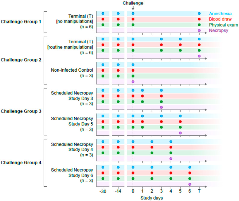
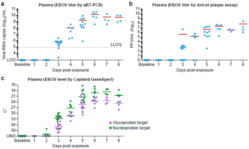
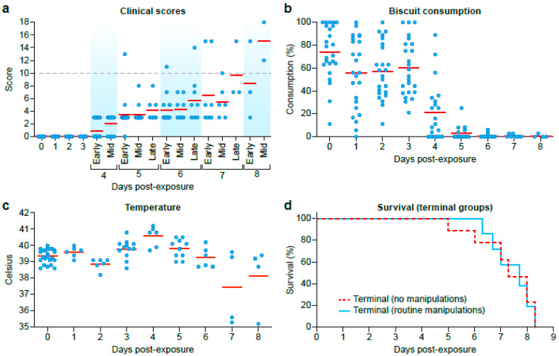
Ongoing Ebola virus disease outbreaks in the Democratic Republic of the Congo follow the largest recorded outbreak in Western Africa (2013-2016). To combat outbreaks, testing of medical countermeasures (therapeutics or vaccines) requires a well-defined, reproducible, animal model. Here we present Ebola virus disease kinetics in 24 Chinese-origin rhesus monkeys exposed intramuscularly to a highly characterized, commercially available Kikwit Ebola virus Filovirus Animal Non-Clinical Group (FANG) stock. Until reaching predetermined clinical disease endpoint criteria, six animals underwent anesthesia for repeated clinical sampling and were compared to six that did not. Groups of three animals were euthanized and necropsied on days 3, 4, 5, and 6 post-exposure, respectively. In addition, three uninfected animals served as controls. Here, we present detailed characterization of clinical and laboratory disease kinetics and complete blood counts, serum chemistries, Ebola virus titers, and disease kinetics for future medical countermeasure (MCM) study design and control data. We measured no statistical difference in hematology, chemistry values, or time to clinical endpoint in animals that were anesthetized for clinical sampling during the acute disease compared to those that were not.
Keywords: BSL-4; Ebola virus; FANG; Kikwit; animal model; emerging pathogens; natural history; rhesus monkey; risk group 4.
Conflict of interest statement
The authors declare no conflict of interest.
Figures
References
-
- Kuhn J.H., Amarasinghe G.K., Perry D.L. Filoviridae. In: Howley P.M., Knipe D.M., Whelan S.P.J., editors. Fields Virology. 7th ed. Wolters Lluwer/Lippincott Williams & Wilkins; Philadelphia, PA, USA: 2020. pp. 449–503.
Publication types
MeSH terms
Grants and funding
LinkOut - more resources
Full Text Sources
Medical
