Review
. 2020 Jul 14;9(7):1687.
doi: 10.3390/cells9071687.

Inflammation in Parkinson's Disease: Mechanisms and Therapeutic Implications

Affiliations
Review

Inflammation in Parkinson's Disease: Mechanisms and Therapeutic Implications

Marta Pajares et al. Cells. .

Abstract

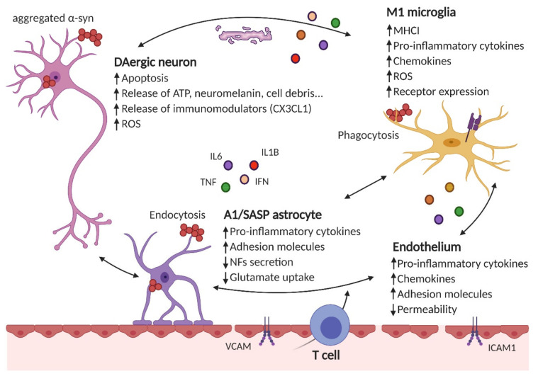
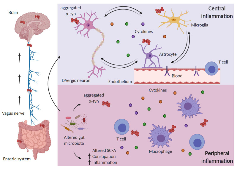
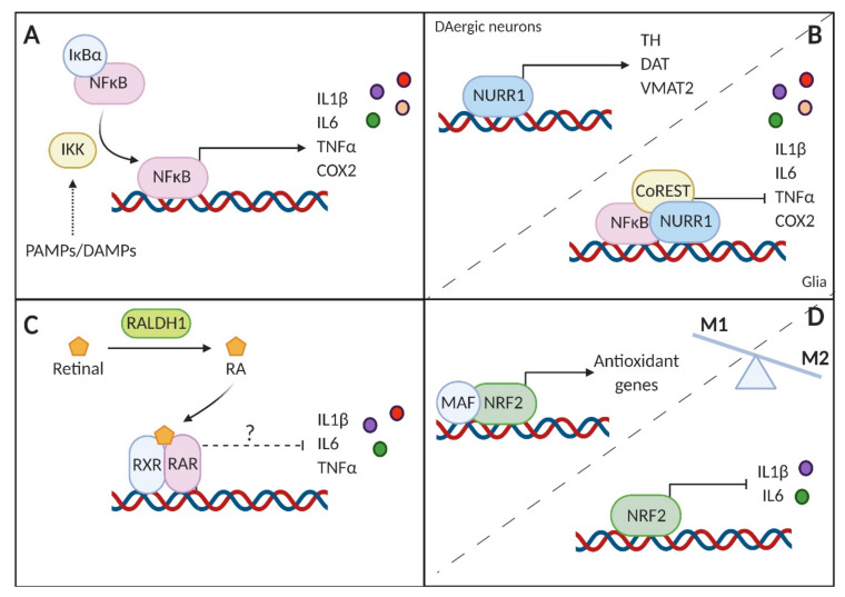
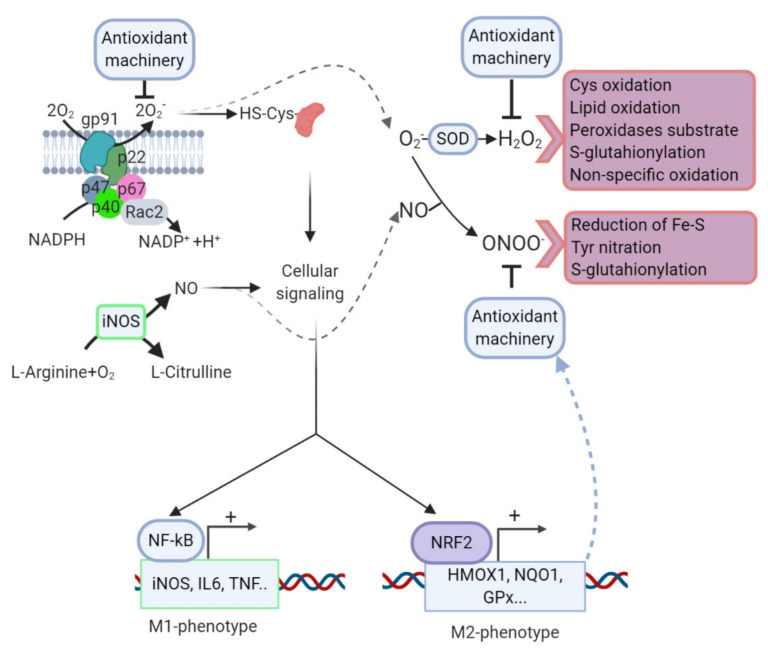
Parkinson's disease (PD) is a common neurodegenerative disorder primarily characterized by the death of dopaminergic neurons that project from the substantia nigra pars compacta. Although the molecular bases for PD development are still little defined, extensive evidence from human samples and animal models support the involvement of inflammation in onset or progression. However, the exact trigger for this response remains unclear. Here, we provide a systematic review of the cellular mediators, i.e., microglia, astroglia and endothelial cells. We also discuss the genetic and transcriptional control of inflammation in PD and the immunomodulatory role of dopamine and reactive oxygen species. Finally, we summarize the preclinical and clinical approaches targeting neuroinflammation in PD.

Keywords: Parkinson’s disease; immune system; neurodegeneration; neuroinflammation; therapy.


Conflict of interest statement

The authors declare that the research was conducted in the absence of any commercial or financial relationships that could be construed as a potential conflict of interest.

Figures

Figure 1
Figure 1
Neuroinflammation results from the crosstalk between different cell types in the brain. Neurons, astrocytes, microglial or endothelial cells are susceptible to α-Syn aggregates (i.e., by phagocytosis, endocytosis, Toll-like receptor (TLR) stimulation, etc.), which can result in the impairment of their homeostatic functions (reduced secretion of neurotrofic factors -NFs-, impaired glutamate uptake, etc.) and secretion of proinflammatory cytokines (such as IL6, IL1β, TNFα, IFNγ, etc.), chemokines (CCL2, CXCL1, etc.) and increased receptor expression (for proinflammatory cytokines and chemokines, MHCI in microglial cells, adhesion molecules in the endothelium, etc.). Additionally, peripheral immune cells (such as CD4+ T cells) are recruited to the brain parenchyma. These immunomodulatory mediators and/or the lack of efficient resolving mechanisms further increase the proinflammatory environment.
Figure 2
Figure 2
Inflammation in Parkinson’s disease (PD) encompasses central and peripheral inflammation. The “gut–brain axis” hypothesis in PD holds that alterations in the gut microbiota may favor α-Syn aggregation and are responsible for an inflammatory response in the periphery, which includes increased cytokine levels and activated T cells. Aggregated α-Syn is suggested to spread from the periphery to the brain through the vagus nerve in a prion-like manner. Once in the brain, proteinopathy together with other triggering factors (mitochondrial impairment, ROS, etc.) will sustain central inflammation in a vicious circle between dying dopaminergic neurons, glial cells and activated endothelium, further aggravated by infiltrating peripheral immune cells.
Figure 3
Figure 3
Schematic representation of the transcriptional modulation of the inflammatory response in PD. (A) Nuclear factor-kappa B (NF-κB) is subjected to tight regulation by the nuclear κ B inhibitor α (IκBα). Upon activation by pathogen-associated or damage-associated molecular patterns (PAMPs or DAMPs, respectively), the IκB kinase (IKK) targets IκBα for degradation, allowing NF-κB to transactivate the expression of proinflammatory genes. (B) Nuclear receptor related 1 protein (NURR1) controls the expression of essential genes for the survival of dopaminergic (DAergic) neurons, but also in glial cells has the potential to repress the activity of NF-κB when recruiting the corepressor complex CoREST. (C) Reduced levels of the enzyme RALDH1 in PD may impair proper synthesis of retinoic acid (RA), essential for the nigrostriatal pathway. Moreover, RA and its derivatives have shown to have key anti-inflammatory effects, that, therefore, would be lessened in PD. (D) NRF2 controls the transcription of antioxidant genes, thereby reducing the oxidative burst. NRF2 directly represses the transcription of the proinflammatory interleukin 1β (IL1β) and interleukin 6 (IL6). The overall effect of this transcription factor is to counteract the proinflammatory phenotype (M1) in favor of the anti-inflammatory (M2) phenotype.
Figure 4
Figure 4
Redox control of microglial phenotype. Under surveillance mode, microglial cells exhibit low levels of ROS/RNS properly managed by the antioxidant machinery. After an inflammatory challenge, microglia activate an M1 program that is characterized through a rapid and high increase in ROS/RNS levels mainly derived from the NOX and NOS activities. During this phase, ROS/RNS act as second messengers increasing the phosphorylation levels of the kinases that control ΝF-κB to further upregulate the proinflammatory M1 gene profile. However, other transcription factors, including NRF2, are increased in response to ROS/RNS but probably in a second wave. NRF2 will restore redox homeostasis and attenuate M1 in favor of M2 phenotypes. Crosstalk between NOX and NOS systems, H2O2 generated after superoxide anion dismutation can induce cysteine oxidation, S-glutathionylation, lipid peroxidation, and a reaction with other peroxides or nonspecific oxidation of other molecules. NO, results of the transformation of L-arginine by NOS, can react with superoxide anion generating peroxynitrate, a molecule that can modify tyrosine residues by nitration, S-glutathionylation of diverse molecules, or reduction of ferric sulfide.

References

    1. Surmeier D.J., Obeso J.A., Halliday G.M. Selective neuronal vulnerability in Parkinson disease. Nat. Rev. Neurosci. 2017;18:101–113. doi: 10.1038/nrn.2016.178. - DOI - PMC - PubMed
    1. Jankovic J., Tan E.K. Parkinson’s disease: Etiopathogenesis and treatment. J. Neurol. Neurosurg. Psychiatry. 2020:322338. doi: 10.1136/jnnp-2019-322338. - DOI - PubMed
    1. Wirdefeldt K., Adami H.O., Cole P., Trichopoulos D., Mandel J. Epidemiology and etiology of Parkinson’s disease: A review of the evidence. Eur. J. Epidemiol. 2011;26(Suppl. 1):S1–S58. doi: 10.1007/s10654-011-9581-6. - DOI - PubMed
    1. Lutters B., Foley P., Koehler P.J. The centennial lesson of encephalitis lethargica. Neurology. 2018;90:563–567. doi: 10.1212/WNL.0000000000005176. - DOI - PubMed
    1. Limphaibool N., Iwanowski P., Holstad M.J.V., Kobylarek D., Kozubski W. Infectious Etiologies of Parkinsonism: Pathomechanisms and Clinical Implications. Front. Neurol. 2019;10:652. doi: 10.3389/fneur.2019.00652. - DOI - PMC - PubMed

Publication types