

Fluorescent Labeling of Helminth Extracellular Vesicles Using an In Vivo Whole Organism Approach
- PMID: 32674418
- PMCID: PMC7399896
- DOI: 10.3390/biomedicines8070213
Fluorescent Labeling of Helminth Extracellular Vesicles Using an In Vivo Whole Organism Approach
Abstract
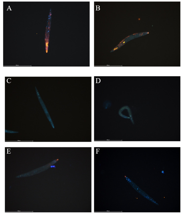
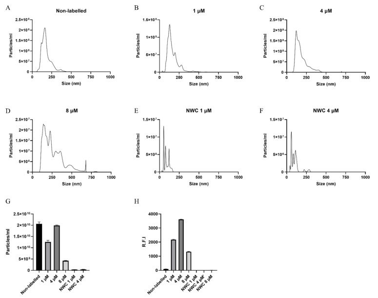
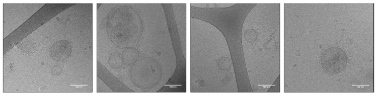
In the last two decades, extracellular vesicles (EVs) from the three domains of life, Archaea, Bacteria and Eukaryotes, have gained increasing scientific attention. As such, the role of EVs in host-pathogen communication and immune modulation are being intensely investigated. Pivotal to EV research is the determination of how and where EVs are taken up by recipient cells and organs in vivo, which requires suitable tracking strategies including labelling. Labelling of EVs is often performed post-isolation which increases risks of non-specific labelling and the introduction of labelling artefacts. Here we exploited the inability of helminths to de novo synthesise fatty acids to enable labelling of EVs by whole organism uptake of fluorescent lipid analogues and the subsequent incorporation in EVs. We showed uptake of 1,2-dioleoyl-sn-glycero-3-phosphoethanolamine-N-(lissamine rhodamine B sulfonyl) (DOPE-Rho) in Anisakis spp. and Trichuris suis larvae. EVs isolated from the supernatant of Anisakis spp. labelled with DOPE-Rho were characterised to assess the effects of labelling on size, structure and fluorescence of EVs. Fluorescent EVs were successfully taken up by the human macrophage cell line THP-1. This study, therefore, presents a novel staining method that can be utilized by the EV field in parasitology and potentially across multiple species.
Keywords: Cryo–EM; extracellular vesicles; helminth; proteomics; vesicle labelling; vesicle tracking.
Conflict of interest statement
The authors declare no conflict of interest. The funders had no role in the design of the study; in the collection, analyses, or interpretation of data; in the writing of the manuscript, or in the decision to publish the results.
Figures


References
-
- Buck A.H., Coakley G., Simbari F., McSorley H.J., Quintana J.F., Le Bihan T., Kumar S., Abreu-Goodger C., Lear M., Harcus Y., et al. Exosomes secreted by nematode parasites transfer small RNAs to mammalian cells and modulate innate immunity. Nat. Commun. 2014;5:1–11. doi: 10.1038/ncomms6488. - DOI - PMC - PubMed
Grants and funding
LinkOut - more resources
Full Text Sources
Research Materials
Miscellaneous
