. 2020 Jul 14;10(7):1195.
doi: 10.3390/ani10071195.

Molecular Characterization and Determination of Relative Cytokine Expression in Naturally Infected Day-Old Chicks with Chicken Astrovirus Associated to White Chick Syndrome

Affiliations

Molecular Characterization and Determination of Relative Cytokine Expression in Naturally Infected Day-Old Chicks with Chicken Astrovirus Associated to White Chick Syndrome

Luis F Naranjo Nuñez et al. Animals (Basel). .

Abstract

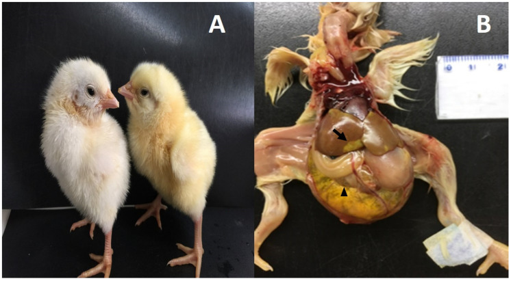
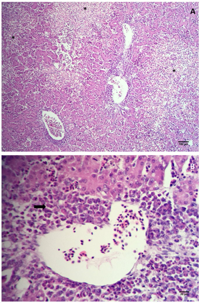
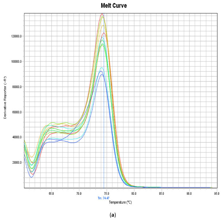
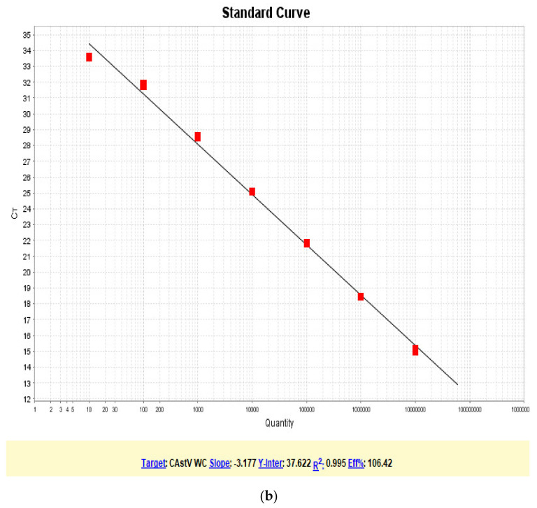
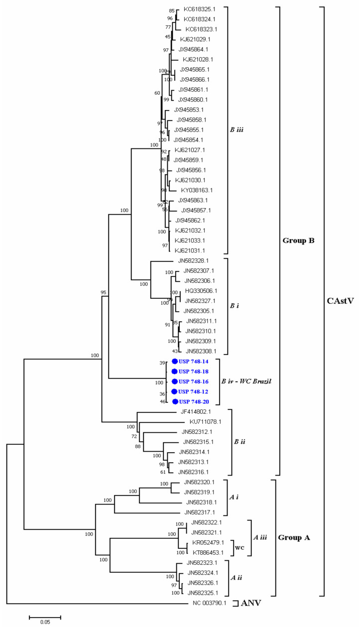
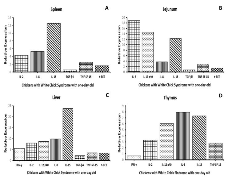
White chick syndrome (WCS) is an emergent disease that affects hatchability and hatched chicks, resulting in high mortality and economic losses, and is related to chicken astrovirus (CAstV). This syndrome has been reported in several countries worldwide, and groups A iii and B vi of CAstV have been determined; however, in Brazil, the virus has not been genotyped. The innate immunity of chicks affected by WCS or any CAstV is poorly understood and studied, and it is important to determine whether relative cytokine expression occurs during the early stages of the life of chicks. The aim of the present investigation is to detect and molecularly characterize CAstV associated with WCS, examine the macroscopic and microscopic lesions in the jejunum and spleen, and determine cytokine expression in the jejunum, liver, spleen and thymus of chicks naturally infected with WCS. To do so, we applied a pathological and molecular approach for CAstV detection and characterization, as well as the quantification of the relative mRNA expression of several cytokine genes. The phylogenetic analyses of the sequences obtained herein classified CAstV as uniquely belonging to group B iv, showing a high similarity of nucleotides (NT) (75.7-80.6%) and amino acids (AA) (84.2-89.9%) with the members of group B and a low similarity of NT (46.7-47.9%) and AA (37.8-38.9%) with the virus belonging in group A. CAstV was also detected and quantified in the serum, spleen, thymus and jejunum, the latter being the organ where CAstV had the highest viral concentration. However, this organ did not present any microscopical alterations. In contrast, we observed necrotic hepatitis in the liver of the affected subjects. On the other hand, we observed the activation of several T helper 1 (Th1)- and T helper 2 (Th2)-cytokines (IFN-γ, IL-2, IL-8, IL-12p40, IL-15, TGF-β4, TNF-SF-15 and t-BET), without being able to control the viral replication due to the high concentration of viral particles in some organs, principally in the gut. One possible role of these cytokines is contributing to the control of inflammation and cell protection of intestinal cells, principally during the early activation of immune responses. However, the fact that these responses are not mature enough to control the viral infection means that more studies need to be carried out to elucidate this topic.

Keywords: Th1; Th2; chicken astrovirus; cytokines; white chicks.


Conflict of interest statement

The authors declare no conflict of interest.

Figures

Figure 1
Figure 1
Macroscopic lesions observed in animals affected with white chick syndrome (WCS); (A): left: chick affected with WCS, right: non-affected chick; (B): macroscopic lesions present in jejunum and liver in the animals with WCS; hepatomegaly, with focal pale yellow areas (arrow), and duodenal loop filled with liquid (arrowhead).
Figure 2
Figure 2
Photomicrography of liver sections from chicks with a one-day-old WCS. Liver parenchyma showed necroses—asterisks (A) and increases in the infiltrate of granulocytes—arrow (B) indicating necrosis hepatitis. All slides were stained with hematoxylin–eosin (HE).
Figure 3
Figure 3
Melting (a) and efficiency (b) curves of the RT-qPCR assay performed to amplify and quantify CAstV associated with WCS. The melting curve showed a melting temperature of 74.47 °C, and the standard curve had an efficiency of 106.4%.
Figure 3
Figure 3
Melting (a) and efficiency (b) curves of the RT-qPCR assay performed to amplify and quantify CAstV associated with WCS. The melting curve showed a melting temperature of 74.47 °C, and the standard curve had an efficiency of 106.4%.
Figure 4
Figure 4
Phylogenetic relations between the sequences of CAstV belonging to chicks naturally infected with WCS obtained in the present work and other sequences of CAstV based on complete ORF2 gene nucleotide sequences. Sequences were aligned using the CLUSTAL W method in ClustaX2 2.1. The phylogenetic tree was constructed using the MEGA 7 Software Package. The numbers along the branches refer to the bootstrap values of 1000 replicates. The scale bar represents the number of substitutions per site. Avian Nephritis virus (ANV) was used as the outgroup. The sequences obtained here are shown in blue.
Figure 5
Figure 5
Relative gene expression of cytokines. Relative gene expression of IFN-γ, IL-2, IL-8, IL-12 p40, IL-15, TGF-β4, TNF-SF-15 and t-BET in the spleen (A), jejunum (B), liver (C) and thymus (D) of chicks naturally affected with white chick syndrome. The data represent the mean gene expression levels of the target genes in five biological replicates relative to the housekeeping gene β-actin.

Similar articles

Cited by

References

    1. Pantin-Jackwood M.J., Spackman E., Woolcock P.R. Molecular characterization and typing of chicken and turkey astroviruses circulating in the United States: Implications for diagnostics. Avian Dis. 2006;50:397–404. doi: 10.1637/7512-020606R.1. - DOI - PubMed
    1. De la Torre D.I.D., Nuñez L.F.L., Astolfi-Ferreira C.S., Ferreira A.J.P. Enteric virus diversity examined by molecular methods in Brazilian poultry flocks. Vet. Sci. 2018;5:38. doi: 10.3390/vetsci5020038. - DOI - PMC - PubMed
    1. Smyth V.J., Todd D., Trudgett J., Lee A., Welsh M.D. Capsid protein sequence diversity of chicken astrovirus. Avian Pathol. 2012;41:151–159. doi: 10.1080/03079457.2011.652938. - DOI - PubMed
    1. Skibinska A., Lee A., Wylie M., Smyth V.J., Welsh M.D., Todd D. Development of an indirect enzyme-linked immunosorbent assay test for detecting antibodies to chicken astrovirus in chicken sera. Avian Pathol. 2015;44:436–442. doi: 10.1080/03079457.2015.1084411. - DOI - PubMed
    1. Xue J., Han T., Xu M., Zhao J., Zhang G. The first serological investigation of Chicken astrovirus infection in China. Biologicals. 2017;47:22–24. doi: 10.1016/j.biologicals.2017.03.005. - DOI - PubMed