Review
. 2020 Jul 14;10(7):449.
doi: 10.3390/brainsci10070449.

Role of NAD+-Modulated Mitochondrial Free Radical Generation in Mechanisms of Acute Brain Injury

Affiliations
Review

Role of NAD+-Modulated Mitochondrial Free Radical Generation in Mechanisms of Acute Brain Injury

Nina Klimova et al. Brain Sci. .

Abstract

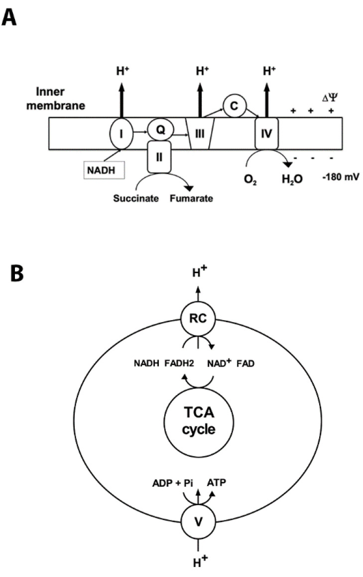
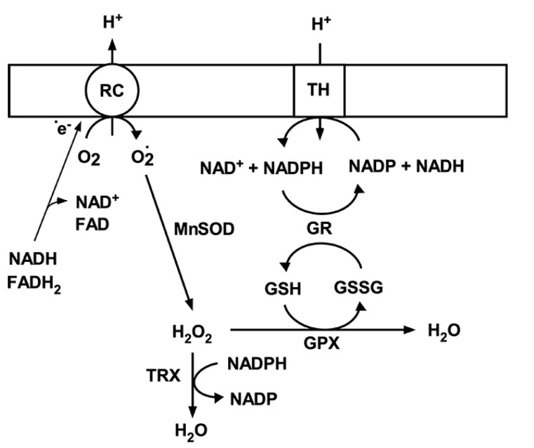
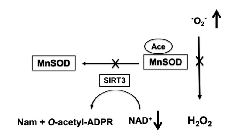
It is commonly accepted that mitochondria represent a major source of free radicals following acute brain injury or during the progression of neurodegenerative diseases. The levels of reactive oxygen species (ROS) in cells are determined by two opposing mechanisms-the one that produces free radicals and the cellular antioxidant system that eliminates ROS. Thus, the balance between the rate of ROS production and the efficiency of the cellular detoxification process determines the levels of harmful reactive oxygen species. Consequently, increase in free radical levels can be a result of higher rates of ROS production or due to the inhibition of the enzymes that participate in the antioxidant mechanisms. The enzymes' activity can be modulated by post-translational modifications that are commonly altered under pathologic conditions. In this review we will discuss the mechanisms of mitochondrial free radical production following ischemic insult, mechanisms that protect mitochondria against free radical damage, and the impact of post-ischemic nicotinamide adenine mononucleotide (NAD+) catabolism on mitochondrial protein acetylation that affects ROS generation and mitochondrial dynamics. We propose a mechanism of mitochondrial free radical generation due to a compromised mitochondrial antioxidant system caused by intra-mitochondrial NAD+ depletion. Finally, the interplay between different mechanisms of mitochondrial ROS generation and potential therapeutic approaches are reviewed.

Keywords: NAD+; acetylation; free radicals; ischemia; mitochondria.


Conflict of interest statement

The authors declare no conflict of interest.

Figures

Figure 1
Figure 1
Mitochondrial oxidative phosphorylation. Oxidative phosphorylation generates ATP using the electrochemical potential (comprised of chemical gradient of hydrogen ions and the membrane potential ∆03A8) formed by the respiratory chain complexes in the inner mitochondrial membrane. (A) Complex I (NADH: ubiquinone oxidoreductase (I)) accepts electrons from NADH, and together with complex II (succinate: ubiquinone oxidoreductase (II)), which oxidizes succinate to fumarate, transfers electrons to ubiquinone (Q). Then the electrons are accepted by complex III (III) and transferred to complex IV (IV) via cytochrome C (C). At complex IV oxygen is reduced to water. Respiratory chain (RC) complexes I, III, and IV pump hydrogen ions (H+) across the inner membrane to generate the protomotive force that drives complex V to synthetize ATP. The thin arrows represent the transfer of the electrons; bold arrows show the hydrogen ions’ transport across the inner membrane. (B) Schematic illustration of coupling between activity of respiratory complexes (RC) and ATP production by complex V (ATP synthase).
Figure 2
Figure 2
Mitochondrial antioxidant system. Superoxide (O2-) generated by respiratory chain complexes (RC) is dismutated to hydrogen peroxide (H2O2) by MnSOD. H2O2 is then converted to water either by glutathione peroxidase using reduced glutathione (GSH) or thioredoxin peroxidase (TRX). Oxidized glutathione (GSSG) is then reduced back to GSH by glutathione peroxidase (GPX) using NADPH as an electron donor. NADP is then reduced by transhydrogenase; that is driven by mitochondrial membrane potential and transferring electrons from NADH.
Figure 3
Figure 3
NAD+-dependent increase in mitochondrial superoxide production (O2-). Ischemia-induced depletion of mitochondrial NAD+ pools leads to reduced activity of SIRT3 that transfers the acetyl group (Ace) from MnSOD to the ADP ribose (ADPR) moiety of NAD+ and forms O-acetyl-ADPR and nicotinamide (Nam). The hyperacetylation-induced inhibition of MnSOD results in insufficient removal of superoxide generated by mitochondria causing an increase in ROS levels.

Similar articles

Cited by

References

    1. Droge W. Free radicals in the physiological control of cell function. Physiol. Rev. 2002;82:47–95. doi: 10.1152/physrev.00018.2001. - DOI - PubMed
    1. Chen Y.R., Zweier J.L. Cardiac mitochondria and reactive oxygen species generation. Circ. Res. 2014;114:524–537. doi: 10.1161/CIRCRESAHA.114.300559. - DOI - PMC - PubMed
    1. Murphy M.P. How mitochondria produce reactive oxygen species. Biochem. J. 2009;417:1–13. doi: 10.1042/BJ20081386. - DOI - PMC - PubMed
    1. Lin M.T., Beal M.F. Mitochondrial dysfunction and oxidative stress in neurodegenerative diseases. Nature. 2006;443:787–795. doi: 10.1038/nature05292. - DOI - PubMed
    1. Tretter L., Takacs K., Hegedus V., Adam-Vizi V. Characteristics of alpha-glycerophosphate-evoked H2O2 generation in brain mitochondria. J. Neurochem. 2007;100:650–663. doi: 10.1111/j.1471-4159.2006.04223.x. - DOI - PubMed

LinkOut - more resources