Case Reports
. 2020 Jul 16;13(7):e234828.
doi: 10.1136/bcr-2020-234828.

Double valve involvement: papillary fibroelastoma in a patient with severe mitral and aortic valve regurgitation

Affiliations
Case Reports

Double valve involvement: papillary fibroelastoma in a patient with severe mitral and aortic valve regurgitation

Oyintayo Ajiboye et al. BMJ Case Rep. .

Abstract

Cardiac papillary fibroelastoma is a benign neoplasm that arises in the endocardium. It commonly presents as an incidental finding on transthoracic echocardiography or as emboli to the coronary, cerebral or pulmonary vasculature. Clinical manifestations described in the literature have generally been related to a sequelae of the associated embolic phenomenon of these lesions. Valve regurgitation is less common with papillary fibroelastoma and when found, it is not known to cause severe regurgitation requiring valve replacement. We report a case of papillary fibroelastoma in a patient with severe mitral and aortic valve regurgitation in association with mobile masses requiring double valve replacement. This patient managed initially as infective endocarditis with severe double valve regurgitation, was found to have valvular masses concernng for papillary fibroelastoma and subsequently confirmed on pathology.

Keywords: cardiothoracic surgery; cardiovascular system; valvar diseases.


Conflict of interest statement

Competing interests: None declared.

Figures

Figure 1
Figure 1
Transthoracic echocardiographic image in parasternal long-axis view showing a 1.6 cm (L)×1.3 cm (W), mobile tumour on the atrial aspect of the tip of the anterior leaflet with ‘sea-anemone’-like appearance, LA, left atrium; LV, left ventricle.
Figure 2
Figure 2
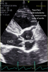
Contrast-enhanced transthoracic echocardiographic image in parasternal long-axis view showing papillary tumour measuring 1.0 cm (L)×0.5 cm (W), with a stalk on the left ventricular aspect of the non-coronary cusp of the aortic valve. AO, aorta; LVOT, left ventricular outflow tract.
Figure 3
Figure 3
Aortic valve (H&E stain) showing the characteristic avascular fronds with endothelial lining of the fibroelastoma.

References

    1. Bruckner BA, Reardon MJ. Benign cardiac tumors: a review. Methodist Debakey Cardiovasc J 2010;6:20–6. 10.14797/mdcj-6-3-20 - DOI - PubMed
    1. Rodrigues JD, Ferreira J, Almeida J, et al. . Cardiac papillary fibroelastoma: report of a surgical series. Rev Port Cardiol 2018;37:981–6. 10.1016/j.repc.2018.02.011 - DOI - PubMed
    1. Logan N, Islam MS, Chughtai JZ, et al. . An atypical cause of myocardial infarction: case report of an obstructing papillary fibroelastoma of the aortic valve. Eur Heart J Case Rep 2019;3:1–3. 10.1093/ehjcr/ytz058 - DOI - PMC - PubMed
    1. Maleszewski JJ, Bois MC, Bois JP, et al. . Neoplasia and the Heart: Pathological Review of Effects With Clinical and Radiological Correlation. J Am Coll Cardiol 2018;72:202–27. 10.1016/j.jacc.2018.05.026 - DOI - PubMed
    1. Wittersheim M, Heydt C, Hoffmann F, et al. . KRAS mutation in papillary fibroelastoma: a true cardiac neoplasm? J Pathol Clin Res 2017;3:100–4. 10.1002/cjp2.66 - DOI - PMC - PubMed

Publication types

MeSH terms