. 2020 Jul;20(4):384-388.
doi: 10.7861/clinmed.2020-0232.

Idiopathic intracranial hypertension: Update on diagnosis and management

Affiliations

Idiopathic intracranial hypertension: Update on diagnosis and management

Benjamin R Wakerley et al. Clin Med (Lond). 2020 Jul.

Abstract

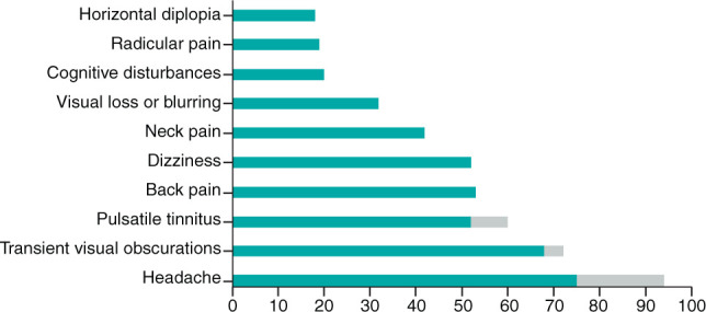
Idiopathic intracranial hypertension is a condition of raised intracranial pressure of unknown cause. Features include new onset headache, which is frequently non-specific; papilloedema is present, visual disturbances are common; and there may be sixth nerve palsy. Diagnosis includes brain imaging with venography to exclude structural causes and venous sinus thrombosis. Lumbar puncture reveals pressure greater than 250 mmCSF with normal constituents. Treatments aim to modify the disease, prevent permanent visual loss and manage headaches. These include weight loss. For those with rapid visual decline, urgent surgical intervention is essential. For most, this is a chronic condition characterised by significantly disabling headaches.

Keywords: Idiopathic intracranial hypertension; headache; obesity; papilloedema; raised intracranial pressure.


Figures

Fig 1.
Fig 1.
Most common symptoms in idiopathic intracranial hypertension.
Fig 2.
Fig 2.
Progressive optical coherence tomography of retinal structures. This patient presented with papilloedema (a–d) was managed locally and re-referred back to our services following weight loss at 14 months with resolving papilloedema (e–h). The images show the difference between colour fundus images (a, c, e, g) and infrared images (b, d, f, h) taken using Heidelberg Spectralis optical coherence tomography. a) The right optic nerve head showing swelling at presentation. b) Greater fidelity image of the swelling of right optic nerve head. c) The left optic nerve head showing swelling at the presentation. d) Comparative image showing left optic nerve head swelling. e) The right optic nerve head showing resolving of the swelling compared with (a). f) Image showing the reduction in swelling compared with (b). g) The left optic nerve head showing resolving of swelling compared with (c). h) Image showing resolving of swelling compared with (d).
Fig 3.
Fig 3.
Scans used to longitudinally monitor papilloedema from the same patient as in Fig 2. a) Infrared (IR) imaging showing the peripapillary retinal nerve fibre layer (RNFL), the green circle shows where data are taken from. b) Resolution over time in the height of the RNFL. c) The volume of the optic nerve head showing changes from the reference scan (top) to the present scan (middle) and the average change in microns (bottom). d) IR imaging and the volume cube scan taken over the optic nerve head. e) The individual line scan for one of the middle slices through the optic nerve head, the fine grey line shows the original extent of the swelling of the right optic nerve, the black shows the present scan and the green band shows the amount of reduction in the swelling.
Fig 4.
Fig 4.
Typical radiological features of stigmata of raised intracranial pressure. a) Axial T2-weighted brain magnetic resonance imaging (MRI) demonstrating widening of the perioptic spaces (arrow), which are normally barely visible in healthy individuals. b) Flattening of the anterior pituitary gland by increased cerebrospinal fluid pressure resulting in a partially empty sella (arrow), which is best appreciated on sagittal T1-weighted brain MRI. c) Left extrinsic compression of the transverse sinus demonstrated here with MRI venography (arrow).

Comment in

  • Idiopathic intracranial hypertension.
    Morrish P. Morrish P. Clin Med (Lond). 2020 Sep;20(5):e138. doi: 10.7861/clinmed.Let.20.5.6. Clin Med (Lond). 2020. PMID: 32934058 Free PMC article. No abstract available.

References

    1. Markey KA, Mollan SP, Jensen RH, Sinclair AJ. Understanding idiopathic intracranial hypertension: mechanisms, management, and future directions. Lancet Neurol 2016;15:78–91. - PubMed
    1. Mollan SP, Aguiar M, Evison F, Frew E, Sinclair AJ. The expanding burden of idiopathic intracranial hypertension. Eye (Lond) 2019;33:478–85. - PMC - PubMed
    1. Hornby C, Mollan SP, Botfield H, O'Reilly MW, Sinclair AJ. Metabolic concepts in idiopathic intracranial hypertension and their potential for therapeutic intervention. J Neuroophthalmol 2018;38:522–30. - PMC - PubMed
    1. O'Reilly MW, Westgate CS, Hornby C, et al. A unique androgen excess signature in idiopathic intracranial hypertension is linked to cerebrospinal fluid dynamics. JCI Insight 2019;4:e125348. - PMC - PubMed
    1. Adderley NJ, Subramanian A, Nirantharakumar K, et al. Association between idiopathic intracranial hypertension and risk of cardiovascular diseases in women in the united kingdom. JAMA Neurol 2019;76:1088–98. - PMC - PubMed