Review
. 2020 Jul 16;133(14):jcs235937.
doi: 10.1242/jcs.235937.

Mitochondrial dynamics during spermatogenesis

Affiliations
Review

Mitochondrial dynamics during spermatogenesis

Grigor Varuzhanyan et al. J Cell Sci. .

Abstract

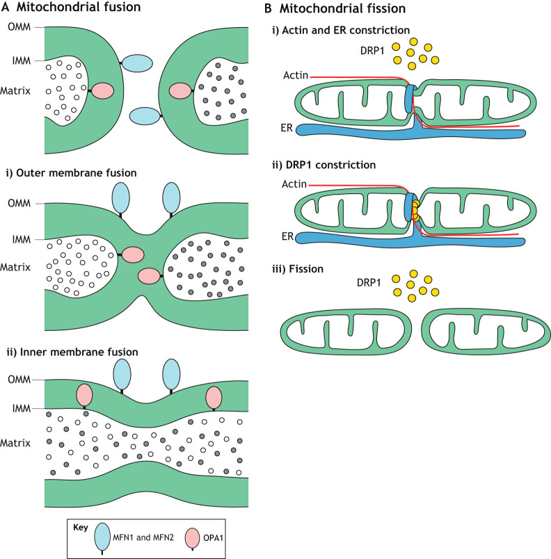
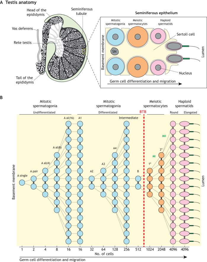
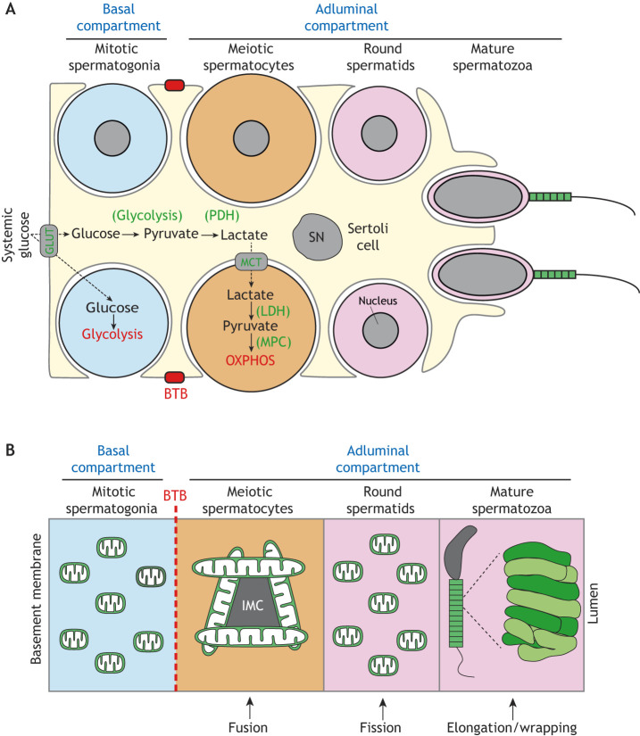
Mitochondrial fusion and fission (mitochondrial dynamics) are homeostatic processes that safeguard normal cellular function. This relationship is especially strong in tissues with constitutively high energy demands, such as brain, heart and skeletal muscle. Less is known about the role of mitochondrial dynamics in developmental systems that involve changes in metabolic function. One such system is spermatogenesis. The first mitochondrial dynamics gene, Fuzzy onions (Fzo), was discovered in 1997 to mediate mitochondrial fusion during Drosophila spermatogenesis. In mammals, however, the role of mitochondrial fusion during spermatogenesis remained unknown for nearly two decades after discovery of Fzo Mammalian spermatogenesis is one of the most complex and lengthy differentiation processes in biology, transforming spermatogonial stem cells into highly specialized sperm cells over a 5-week period. This elaborate differentiation process requires several developmentally regulated mitochondrial and metabolic transitions, making it an attractive model system for studying mitochondrial dynamics in vivo We review the emerging role of mitochondrial biology, and especially its dynamics, during the development of the male germ line.

Keywords: Membrane fission; Membrane fusion; Mitochondrial dynamics; Spermatogenesis.


Conflict of interest statement

Competing interestsThe authors declare no competing or financial interests.

Figures

Fig. 1.
Fig. 1.
Mitochondrial fusion and fission. (A) Mitochondrial fusion occurs in two distinct steps, both mediated by large GTP-hydrolyzing enzymes of the dynamin superfamily. MFN1 and MFN2 mediate fusion of the mitochondrial outer membrane (OMM). Then, OPA1 mediates fusion of the inner membrane (IMM), which results in mixing of matrix components. Although OPA1 is present on opposing IMMs, it is not required to be present on both membranes. (B) Mitochondrial fission is a multistep process. In the initial phase, actin and the ER associate with the mitochondrial tubule. The ER wraps around and constricts the mitochondrion. Receptors on the mitochondrial outer membrane (not shown) recruit cytosolic DRP1 to this constriction site. Multiple DRP1 molecules oligomerize around the mitochondrion to form a ring-shaped structure that further constricts and severs the mitochondrial tubule.
Fig. 2.
Fig. 2.
Mitophagy. (A) Overview of mitophagy. An autophagosome engulfs a damaged portion of a mitochondrion to form a mitophagosome that fuses with lysosomes. The mitochondrion is degraded in the resulting mitolysosome. (B) A model showing the role of FIS1 during PINK1-mediated mitophagy. FIS1 at the damaged mitochondrial surface interacts with TBC1D15, a mitochondrial Rab GAP that inactivates RAB7A, to regulate mitophagosome formation. (C) In the absence of FIS1, the uncontrolled action of RAB7A disrupts autophagosome membrane dynamics, resulting in aberrant mitophagy.
Fig. 3.
Fig. 3.
Spermatogenesis. (A) Left panel, anatomy of the mammalian testis highlighting the convoluted seminiferous tubules in which spermatogenesis takes place. Right panel, schematic illustration of the seminiferous epithelium highlighting the intimate association between somatic Sertoli cells and germ cells. For simplicity, only the major germ cell types are shown. (B) Cellular pedigree of a single undifferentiated spermatogonium, highlighting germ cell amplification. The theoretical number of syncytial cells at each stage is shown at the bottom. Note that meiotic spermatocytes and post-meiotic spermatids develop on the adluminal side of the blood-testis barrier (BTB). SN, Sertoli cell nucleus; A al, A aligned; 1°, primary spermatocyte; 2°, secondary spermatocyte; MI, meiosis I; MII, meiosis II.
Fig. 4.
Fig. 4.
Mitochondrial respiration and dynamics during spermatogenesis. (A) Mitochondrial respiration during spermatogenesis. Spermatogonia in the basal compartment have direct access to systemic glucose, which they use for glycolysis. Spermatocytes and spermatids in the adluminal compartment, however, are separated from the vasculature and interstitial space by the BTB, and thus rely on Sertoli cells for a carbon source. Sertoli cells take up systemic glucose via glucose transporters (GLUTs) and glycolytically convert it into pyruvate, which is converted into lactate via pyruvate dehydrogenase (PDH). Lactate is then shuttled by monocarboxylate transporters (MCT) into spermatocytes, which convert it back into pyruvate via lactate dehydrogenase (LDH). Finally, pyruvate is imported into mitochondria by the mitochondrial pyruvate carrier (MPC) for fueling oxidative phosphorylation (OXPHOS). The nuclei of the lower spermatogonium and spermatocyte are omitted for clarity. (B) Mitochondrial dynamics during spermatogenesis. Mitochondria are generally small and spherical in spermatogonia, which reside in the basal compartment. Upon traversing the BTB (dashed red line) and entering the adluminal compartment, mitochondria elongate and cluster around the nuage, also referred to as intermitochondrial cement (IMC). In post-meiotic spermatids, mitochondria fragment. Finally, near the end of spermiogenesis, mitochondria elongate and tightly pack around the sperm midpiece. SN, Sertoli cell nucleus.
Fig. 5.
Fig. 5.
Mitochondrial reorganization during spermiogenesis. Schematic of spermiogenesis highlighting the formation of the acrosome and the reorganization of mitochondria. During spermatid elongation, a subset of mitochondria line the sperm midpiece, while the rest are transferred into residual bodies for phagocytic degradation in Sertoli cells. It is unknown how the cell determines the fate of these two mitochondrial populations. The acrosome (blue) is another organelle that undergoes drastic reorganization during spermiogenesis.

Similar articles

Cited by

References

    1. Aloisio G. M., Nakada Y., Saatcioglu H. D., Peña C. G., Baker M. D., Tarnawa E. D., Mukherjee J., Manjunath H., Bugde A., Sengupta A. L. et al. (2014). PAX7 expression defines germline stem cells in the adult testis. J. Clin. Invest. 124, 3929-3944. 10.1172/JCI75943 - DOI - PMC - PubMed
    1. Bae J.-W., Kim S.-H., Kim D.-H., Ha J. J., Yi J. K., Hwang S., Ryu B.-Y., Pang M.-G. and Kwon W.-S. (2019). Ras-related proteins (Rab) are key proteins related to male fertility following a unique activation mechanism. Reprod. Biol. 19, 356-362. 10.1016/j.repbio.2019.10.001 - DOI - PubMed
    1. Bajpai M., Gupta G. and Setty B. S. (1998). Changes in carbohydrate metabolism of testicular germ cells during meiosis in the rat. Eur. J. Endocrinol. 138, 322-327. 10.1530/eje.0.1380322 - DOI - PubMed
    1. Baklouti-Gargouri S., Ghorbel M., Ben Mahmoud A., Mkaouar-Rebai E., Cherif M., Chakroun N., Sellami A., Fakhfakh F. and Ammar-Keskes L. (2014). Identification of a novel m.9588G>a missense mutation in the mitochondrial COIII gene in asthenozoospermic Tunisian infertile men. J. Assist. Reprod. Genet. 31, 595-600. 10.1007/s10815-014-0187-2 - DOI - PMC - PubMed
    1. Bao J. and Bedford M. T. (2016). Epigenetic regulation of the histone-to-protamine transition during spermiogenesis. Reproduction 151, R55-R70. 10.1530/REP-15-0562 - DOI - PMC - PubMed

Publication types

Substances

LinkOut - more resources