Case Reports
. 2020 May 4;33(3):430-432.
doi: 10.1080/08998280.2020.1755199. eCollection 2020 Jul.

Optimum treatment for primary intracranial Ewing sarcoma

Affiliations
Case Reports

Optimum treatment for primary intracranial Ewing sarcoma

Sasha G Howell et al. Proc (Bayl Univ Med Cent). .

Abstract

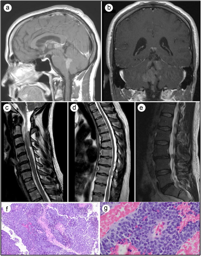
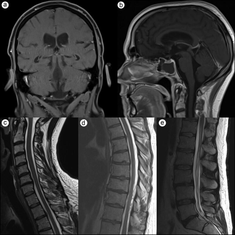
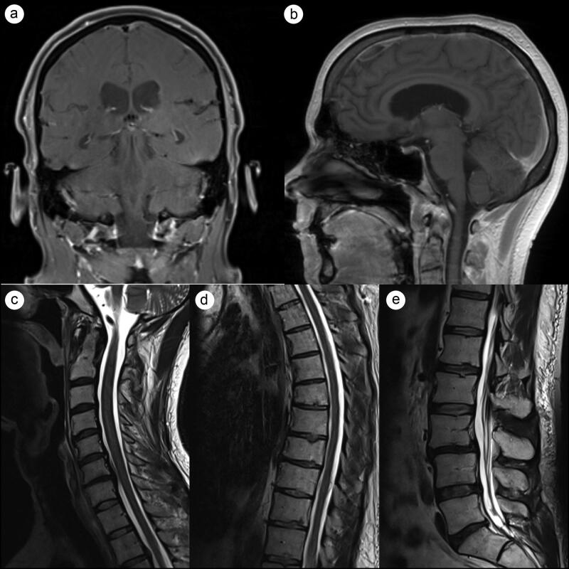
Ewing sarcoma (ES) is an aggressive, high-grade neuroectodermal neoplasm that frequently manifests in children and young adults. Although ES without osseous involvement most commonly involves paravertebral regions of the spine, it very rarely presents as a primary intracranial tumor. This report discusses a unique presentation of an adult extraosseous ES arising from the pineal region with extension into the third and fourth ventricles and multiple drop metastases to the spine. This case demonstrates the application of current chemotherapeutic and adjuvant management and offers insight into possible treatment modalities for metastasis in an atypical extraosseous ES involving the brain and spine.

Keywords: Chemotherapy; Ewing sarcoma; pineal region tumor; radiation therapy.


Figures

Figure 1.
Figure 1.
Pretreatment MRI and histology findings. (a,b) T1-weighted brain MRI with contrast showing multiple homogenous enhancing masses of the third ventricle and posterior fossa. (c,d,e) T2-weighted MRI of cervical, thoracic, and lumbar spines, respectively, showing multiple enhancing intradural lesions at various levels. (f,g) Histopathologic features of Ewing sarcoma/peripheral primitive neuroectodermal tumor. Hematoxylin and eosin staining revealed various sheets of small round blue cells with an increased nuclear to cytoplasmic ratio and high mitotic activity.
Figure 2.
Figure 2.
Three-month follow-up T1-weighted brain MRI and T2-weighted spine MRI. (a,b) Marked tumor regression with no residual intracranial or (c) cervical enhancing lesions. (d) One area of minimal enhancement remained in the thoracic spine. (e) The lumbar spine was significantly improved with only minor residual thickening of the cauda equina.
Figure 3.
Figure 3.
Seven-month follow-up T1-weighted brain MRI and T2-weighted spine MRI. (a,b) Stable tumor regression with no residual intracranial lesions. (c-e) Marked regression of metastatic spinal lesions with no enhancement in (d) thoracic or (e) lumbar spine.

References

    1. Louis DN, Ohgaki H, Wiestler OD, et al. . The 2007 WHO classification of tumours of the central nervous system. Acta Neuropathol. 2007;114(2):97–109. doi:10.1007/s00401-007-0243-4. - DOI - PMC - PubMed
    1. Louis DN, Ohgaki H, Wiestler OD, Cavenee WK. WHO Classification of Tumours of the Central Nervous System. 4th ed. Lyon, France: IARC Press; 2016.
    1. Ladanyi M, Lewis R, Garin-Chesa P, et al. . EWS rearrangement in Ewing’s sarcoma and peripheral neuroectodermal tumor. Molecular detection and correlation with cytogenetic analysis and MIC2 expression. Diagn Mol Pathol. 1993;2:141–146. - PubMed
    1. Pekala JS, Gururangan S, Provenzale JM, et al. . Central nervous system extraosseous Ewing sarcoma: radiologic manifestations of this newly defined pathologic entity. AJNR Am J Neuroradiol. 2006;27(3):580–583. - PMC - PubMed
    1. Jay V, Zielenska M, Lorenzana A, Drake J. An unusual cerebellar primitive neuroectodermal tumor with t(11;22) translocation: pathological and molecular analysis. Fetal & Pediatric Path. 1996;16(1):119–128. doi:10.3109/15513819609168668. - DOI - PubMed

Publication types

LinkOut - more resources