

A 3D biofabricated cutaneous squamous cell carcinoma tissue model with multi-channel confocal microscopy imaging biomarkers to quantify antitumor effects of chemotherapeutics in tissue
- PMID: 32676161
- PMCID: PMC7343636
- DOI: 10.18632/oncotarget.27570
A 3D biofabricated cutaneous squamous cell carcinoma tissue model with multi-channel confocal microscopy imaging biomarkers to quantify antitumor effects of chemotherapeutics in tissue
Abstract
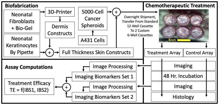
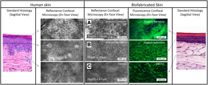
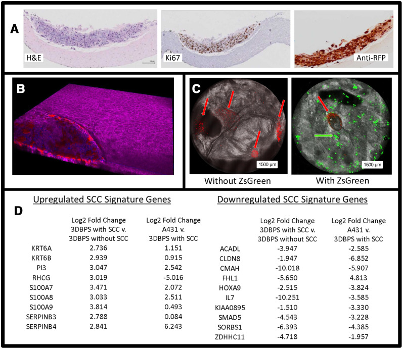
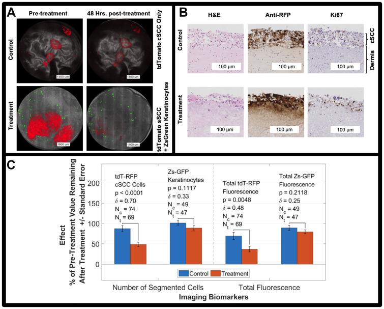
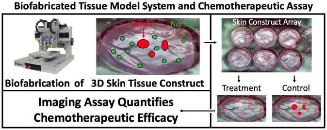
Cutaneous squamous cell carcinoma (cSCC) causes approximately 10,000 deaths annually in the U. S. Current therapies are largely ineffective against metastatic and locally advanced cSCC. There is a need to identify novel, effective, and less toxic small molecule cSCC therapeutics. We developed a 3-dimensional bioprinted skin (3DBPS) model of cSCC tumors together with a microscopy assay to test chemotherapeutic effects in tissue. The full thickness SCC tissue model was validated using hematoxylin and eosin (H&E) and immunohistochemical histological staining, confocal microscopy, and cDNA microarray analysis. A nondestructive, 3D fluorescence confocal imaging assay with tdTomato-labeled A431 SCC and ZsGreen-labeled keratinocytes was developed to test efficacy and general toxicity of chemotherapeutics. Fluorescence-derived imaging biomarkers indicated that 50% of cancer cells were killed in the tissue after 1μM 5-Fluorouracil 48-hour treatment, compared to a baseline of 12% for untreated controls. The imaging biomarkers also showed that normal keratinocytes were less affected by treatment (11% killed) than the untreated tissue, which had no significant killing effect. Data showed that 5-Fluorouracil selectively killed cSCC cells more than keratinocytes. Our 3DBPS assay platform provides cellular-level measurement of cell viability and can be adapted to achieve nondestructive high-throughput screening (HTS) in bio-fabricated tissues.
Keywords: 3D printing; confocal microscopy; in vitro model; screening; squamous cell carcinoma.
Copyright: © 2020 Browning et al.
Conflict of interest statement
CONFLICTS OF INTEREST The authors state no conflicts of interest.
Figures
References
-
- GBD 2015 Disease and Injury Incidence and Prevalence Collaborators Global, regional, and national incidence, prevalence, and years lived with disability for 310 diseases and injuries, 1990-2015: a systematic analysis for the Global Burden of Disease Study 2015. Lancet. 2016; 388:1545–602. 10.1016/S0140-6736(16)31678-6. - DOI - PMC - PubMed
-
- Stratigos A, Garbe C, Lebbe C, Malvehy J, del Marmol V, Pehamberger H, Peris K, Becker JC, Zalaudek I, Saiag P, Middleton MR, Bastholt L, Testori A, Grob JJ, and European Dermatology Forum (EDF), and European Association of Dermato-Oncology (EADO), and European Organization for Research and Treatment of Cancer (EORTC) . Diagnosis and treatment of invasive squamous cell carcinoma of the skin: european consensus-based interdisciplinary guideline. Eur J Cancer. 2015; 51:1989–2007. 10.1016/j.ejca.2015.06.110. - DOI - PubMed
Grants and funding
LinkOut - more resources
Full Text Sources
Research Materials
