[Preprint]. 2020 Jul 6:2020.07.04.20142752.
doi: 10.1101/2020.07.04.20142752.

Mapping Systemic Inflammation and Antibody Responses in Multisystem Inflammatory Syndrome in Children (MIS-C)

Affiliations

Mapping Systemic Inflammation and Antibody Responses in Multisystem Inflammatory Syndrome in Children (MIS-C)

Conor Gruber et al. medRxiv. .

Update in

  • Mapping Systemic Inflammation and Antibody Responses in Multisystem Inflammatory Syndrome in Children (MIS-C).
    Gruber CN, Patel RS, Trachtman R, Lepow L, Amanat F, Krammer F, Wilson KM, Onel K, Geanon D, Tuballes K, Patel M, Mouskas K, O'Donnell T, Merritt E, Simons NW, Barcessat V, Del Valle DM, Udondem S, Kang G, Gangadharan S, Ofori-Amanfo G, Laserson U, Rahman A, Kim-Schulze S, Charney AW, Gnjatic S, Gelb BD, Merad M, Bogunovic D. Gruber CN, et al. Cell. 2020 Nov 12;183(4):982-995.e14. doi: 10.1016/j.cell.2020.09.034. Epub 2020 Sep 14. Cell. 2020. PMID: 32991843 Free PMC article.

Abstract

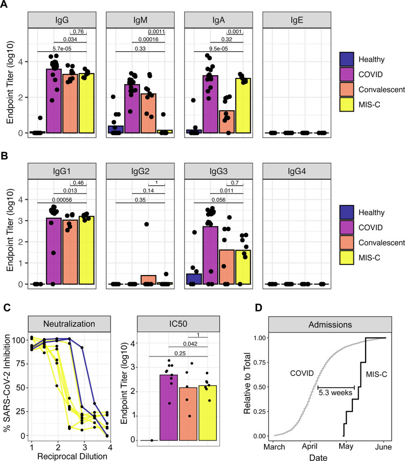
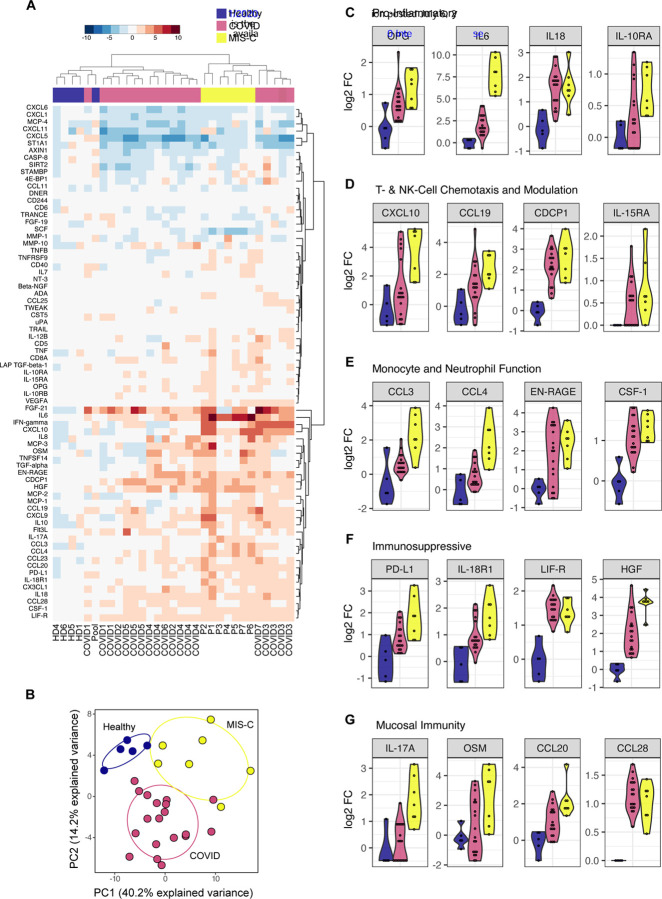
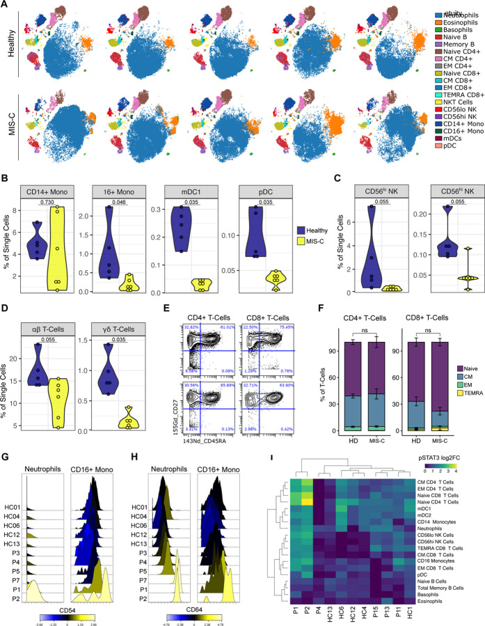
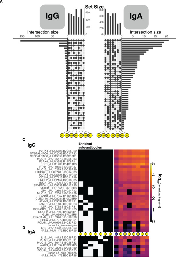
Initially, the global outbreak of COVID-19 caused by severe acute respiratory syndrome coronavirus 2 (SARS-CoV-2) spared children from severe disease. However, after the initial wave of infections, clusters of a novel hyperinflammatory disease have been reported in regions with ongoing SARS-CoV-2 epidemics. While the characteristic clinical features are becoming clear, the pathophysiology remains unknown. Herein, we report on the immune profiles of eight Multisystem Inflammatory Syndrome in Children (MIS-C) cases. We document that all MIS-C patients had evidence of prior SARS-CoV-2 exposure, mounting an antibody response with normal isotype-switching and neutralization capability. We further profiled the secreted immune response by high-dimensional cytokine assays, which identified elevated signatures of inflammation (IL-18 and IL-6), lymphocytic and myeloid chemotaxis and activation (CCL3, CCL4, and CDCP1) and mucosal immune dysregulation (IL-17A, CCL20, CCL28). Mass cytometry immunophenotyping of peripheral blood revealed reductions of mDC1 and non-classical monocytes, as well as both NK- and T- lymphocytes, suggesting extravasation to affected tissues. Markers of activated myeloid function were also evident, including upregulation of ICAM1 and FcγR1 in neutrophil and non-classical monocytes, well-documented markers in autoinflammation and autoimmunity that indicate enhanced antigen presentation and Fc-mediated responses. Finally, to assess the role for autoimmunity secondary to infection, we profiled the auto-antigen reactivity of MIS-C plasma, which revealed both known disease-associated autoantibodies (anti-La) and novel candidates that recognize endothelial, gastrointestinal and immune-cell antigens. All patients were treated with anti-IL6R antibody or IVIG, which led to rapid disease resolution tracking with normalization of inflammatory markers.

Keywords: COVID19; Kawasaki-like; MIS-C; PIMS; SARS-CoV-2; autoimmunity; dysfunction; immune; pediatrics.


Conflict of interest statement

Declaration of Interests DB reports ownership in Lab11 Therapeutics. S.G. reports consultancy and/or advisory roles for Merck, Neon Therapeutics and OncoMed and research funding from Bristol-Myers Squibb, Genentech, Immune Design, Agenus, Janssen R&D, Pfizer, Takeda, and Regeneron.

Figures

Figure 1.
Figure 1.
Anti-SARS-CoV-2 humoral response in MIS-C patients. (A) Endpoint titers against SARS-CoV-2 spike protein in plasma from healthy donors, patients hospitalized for acute COVID-19, convalescent individuals, and MIS-C patients. (B) IgG subtypes. (C) Left: Neutralization of SARS-CoV-2 in Vero cells by plasma from MIS-C patients or a representative COVID positive control. Right: IC50 values for neutralization curves across the full dataset. (D) Admissions for COVID-19 and MIS-C, expressed as a relative proportion of total cases, respectively. Time indicates the delay between COVID-19 and MIS-C in the date of 50% total caseload.
Figure 2.
Figure 2.
Cytokine profiling of plasma from MIS-C and pediatric COVID-19 patients. (A) Multiplex cytokine analysis by O-link ELISA, expressed as log2 fold-change over the mean healthy controls. (B). Principal component analysis. (C-G) Cytokines reaching statistical significance in MIS-C samples over healthy controls.
Figure 3.
Figure 3.
Immunophenotyping of MIS-C patient peripheral blood by mass cytometry. (A) Representative tSNE plots demonstrating the immune cell distribution in whole blood from age-matched healthy controls and MIS-C patients. (B) Monocyte and dendritic cell sub-population frequencies quantified as percent of single cells. (C) NK-cell subsets. (D) T-Cell subsets. (E) Representative scatterplots for naive, central memory (CM), effector memory (EM), and T Effector Memory re-expressing CD45RA (TEMRA) cells in a representative healthy donor (top) and MIS-C patient (bottom). (F) Quantification of T-cell subsets across samples. (G) CD54 and (H) CD64 expression in neutrophils and CD16+ monocytes, color-coded by the log2FC over healthy donor median signal intensity. (I) STAT3 phosphorylation across immune cell subtypes, expressed as log2FC. Abbreviations
Figure 4.
Figure 4.
Autoantibody detection unveils an autoreactive repertoire enriched in MIS-C patients. (A-B) Upset plots delineating the number of shared autoantibodies which were at least two-fold enriched when compared to controls for IgG and IgA. (C-D) Tile plots and heatmaps showing the presence or not, and quantile normalized quantity of IgG and IgA autoantigen reactive antibodies that were at least four-fold enriched when compared to controls and found in 4 patients or more, inclusive of patient P3.

References

    1. Amanat F., Stadlbauer D., Strohmeier S., Nguyen T.H.O., Chromikova V., McMahon M., Jiang K., Arunkumar G.A., Jurczyszak D., Polanco J., et al. (2020). A serological assay to detect SARS-CoV-2 seroconversion in humans. Nat. Med. - PMC - PubMed
    1. Chang L.-Y., Lu C.-Y., Shao P.-L., Lee P.-I., Lin M.-T., Fan T.-Y., Cheng A.-L., Lee W.-L., Hu J.-J., Yeh S.-J., et al. (2014). Viral infections associated with Kawasaki disease. J. Formos. Med. Assoc. 113, 148–154. - PMC - PubMed
    1. Cheung E.W., Zachariah P., Gorelik M., Boneparth A., Kernie S.G., Orange J.S., and Milner J.D. (2020). Multisystem Inflammatory Syndrome Related to COVID-19 in Previously Healthy Children and Adolescents in New York City. JAMA 8–10. - PMC - PubMed
    1. Dietz S.M., van Stijn D., Burgner D., Levin M., Kuipers I.M., Hutten B.A., and Kuijpers T.W. (2017). Dissecting Kawasaki disease: a state-of-the-art review. Eur. J. Pediatr. 176, 995–1009. - PMC - PubMed
    1. DiMaggio C., Klein M., Berry C., and Frangos S. (2020). Blacks/African Americans are 5 Times More Likely to Develop COVID-19: Spatial Modeling of New York City ZIP Code-level Testing Results. MedRxiv 2020.05.14.20101691. - PMC - PubMed

Publication types