

Characterizing placental stiffness using ultrasound shear-wave elastography in healthy and preeclamptic pregnancies
- PMID: 32676857
- PMCID: PMC7646518
- DOI: 10.1007/s00404-020-05697-x
Characterizing placental stiffness using ultrasound shear-wave elastography in healthy and preeclamptic pregnancies
Abstract
Purpose: To measure the stiffness of the placenta in healthy and preeclamptic patients in the second and third trimesters of pregnancy using ultrasound shear-wave elastography (SWE). We also aimed to evaluate the effect of age, gestational age, gravidity, parity and body mass index (BMI) on placental stiffness and a possible correlation of stiffness with perinatal outcomes.
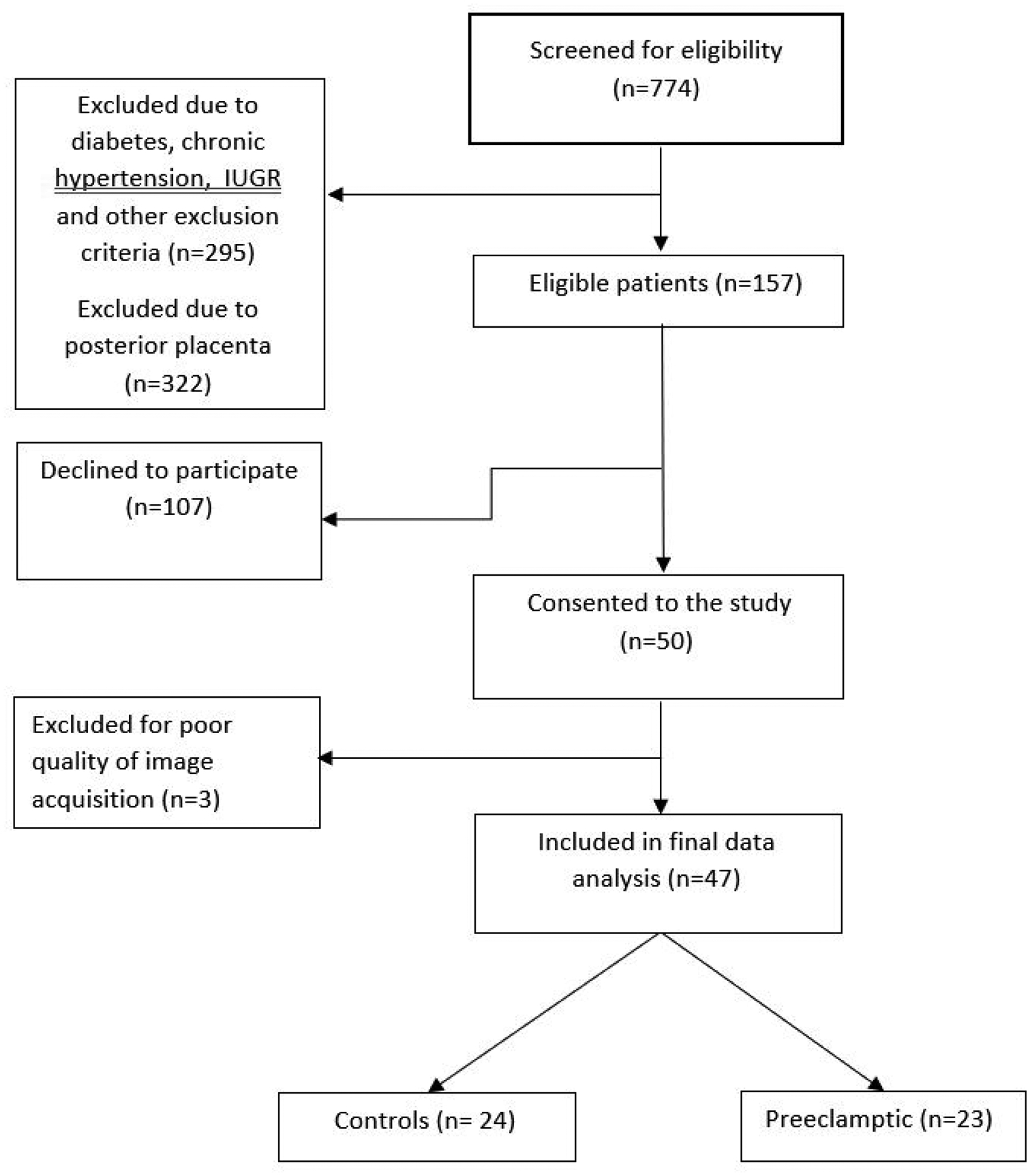
Methods: In a case-control study, we recruited a total of 47 singleton pregnancies in the second and third trimesters of which 24 were healthy and 23 were diagnosed with preeclampsia. In vivo placental stiffness was measured once at the time of recruitment for each patient. Pregnancies with posterior placentas, multiple gestation, gestational hypertension, chronic hypertension, diabetes, autoimmune disease, fetal growth restriction and congenital anomalies were excluded.
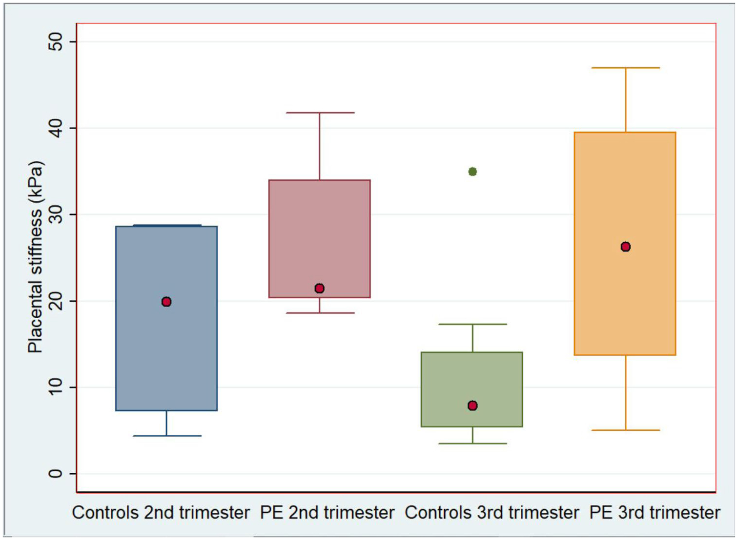
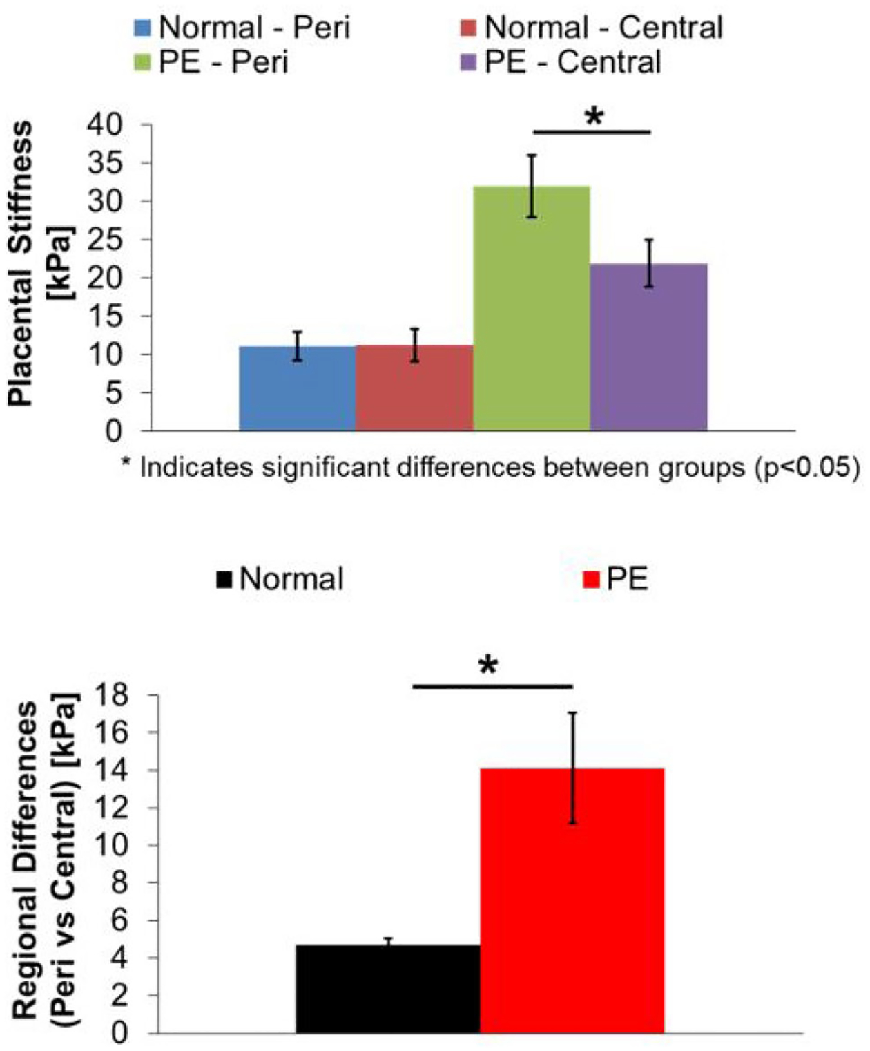
Results: The mean placental stiffness was significantly higher in preeclamptic pregnancies compared to controls in the third trimester (difference of means = 16.8; 95% CI (9.0, 24.5); P < 0.001). There were no significant differences in placental stiffness between the two groups in the second trimester or between the severe preeclampsia and preeclampsia without severe features groups (difference of means = 9.86; 95% CI (-5.95, 25.7); P ≥ 0.05). Peripheral regions of the placenta were significantly stiffer than central regions in the preeclamptic group (difference of means = 10.67; 95% CI (0.07, 21.27); P < 0.05), which was not observed in the control group (difference of means = 0.55; 95% CI (- 5.25, 6.35); P > 0.05). We did not identify a correlation of placental stiffness with gestational age, maternal age, gravidity or parity. However, there was a statistically significant correlation with BMI (P < 0.05). In addition, pregnancies with higher placental stiffness during the 2nd and 3rd trimesters had significantly reduced birth weight (2890 ± 176 vs. 2420 ± 219 g) and earlier GA (37.8 ± 0.84 vs. 34.3 ± 0.98 weeks) at delivery (P < 0.05).
Conclusion: Compared to healthy pregnancies, placentas of preeclamptic pregnancies are stiffer and more heterogeneous. Placental stiffness is not affected by gestational age or the severity of preeclampsia but there is a correlation with higher BMI and poor perinatal outcomes.
Keywords: In vivo; Placenta; Preeclampsia; Shear-wave elastography; Ultrasound.
Conflict of interest statement
Figures


Similar articles
-
Value of shear-wave elastography and cerebral-placental-uterine ratio in women diagnosed with preeclampsia and fetal growth restriction in prediction of adverse perinatal outcomes.J Matern Fetal Neonatal Med. 2022 Dec;35(25):10001-10009. doi: 10.1080/14767058.2022.2081804. Epub 2022 Jun 1. J Matern Fetal Neonatal Med. 2022. PMID: 35647897
-
Shear wave elastography of placenta: in vivo quantitation of placental elasticity in preeclampsia.Diagn Interv Radiol. 2015 May-Jun;21(3):202-7. doi: 10.5152/dir.2014.14338. Diagn Interv Radiol. 2015. PMID: 25858523 Free PMC article.
-
Shear wave elastography in placental dysfunction: comparison of elasticity values in normal and preeclamptic pregnancies in the second trimester.J Ultrasound Med. 2015 Jan;34(1):151-9. doi: 10.7863/ultra.34.1.151. J Ultrasound Med. 2015. PMID: 25542951
-
Placental abnormalities in type 1 and type 2 diabetes mellitus: a systematic review and metaanalysis of shear wave elastography.Am J Obstet Gynecol MFM. 2022 Nov;4(6):100736. doi: 10.1016/j.ajogmf.2022.100736. Epub 2022 Aug 30. Am J Obstet Gynecol MFM. 2022. PMID: 36049626 Review.
-
Systematic review and meta-analysis: gray-scale ultrasound and shear wave elastography in the diagnosis of primipara pregnancy and delivery.Ann Palliat Med. 2021 Nov;10(11):11664-11677. doi: 10.21037/apm-21-2672. Ann Palliat Med. 2021. PMID: 34872291
Cited by
-
Detection of placental stiffness using virtual magnetic resonance elastography in pregnancies complicated by preeclampsia.Arch Gynecol Obstet. 2024 Oct;310(4):2283-2289. doi: 10.1007/s00404-024-07585-0. Epub 2024 Jun 17. Arch Gynecol Obstet. 2024. PMID: 38884644 Free PMC article. No abstract available.
-
Longitudinal associations between urinary biomarkers of phthalates and replacements with novel in vivo measures of placental health.Hum Reprod. 2024 Sep 1;39(9):2104-2114. doi: 10.1093/humrep/deae152. Hum Reprod. 2024. PMID: 38970902 Free PMC article.
-
Micro-RNAs in Human Placenta: Tiny Molecules, Immense Power.Molecules. 2022 Sep 13;27(18):5943. doi: 10.3390/molecules27185943. Molecules. 2022. PMID: 36144676 Free PMC article. Review.
-
The Value of Ultrasonic Elastography in Detecting Placental Stiffness for the Diagnosis of Preeclampsia: A Meta-Analysis.Diagnostics (Basel). 2023 Sep 9;13(18):2894. doi: 10.3390/diagnostics13182894. Diagnostics (Basel). 2023. PMID: 37761261 Free PMC article. Review.
-
A straightforward cell culture insert model to incorporate biochemical and biophysical stromal properties into transplacental transport studies.bioRxiv [Preprint]. 2024 Apr 25:2024.04.19.590317. doi: 10.1101/2024.04.19.590317. bioRxiv. 2024. Update in: Placenta. 2025 Jun 13;166:54-61. doi: 10.1016/j.placenta.2024.09.001. PMID: 38712271 Free PMC article. Updated. Preprint.
References
-
- Bercoff J, Tanter M, Fink M (2004) Supersonic shear imaging: a new technique for soft tissue elasticity mapping. IEEE Trans Ultrason Ferroelectr Freq Control 51(4):396–409 - PubMed
-
- Gennisson J-L, Deffieux T, Fink M, Tanter M (2013) Ultrasound elastography: principles and techniques. Diagn Interv Imaging 94(5):487–495 - PubMed
-
- Sebag F, Vaillant-Lombard J, Berbis J, Griset V, Henry JF, Petit P et al. (2010) shear-wave elastography: a new ultrasound imaging mode for the differential diagnosis of benign and malignant thyroid nodules. J Clin Endocrinol Metab 95(12):5281–5288 - PubMed
-
- Gennisson J-L, Deffieux T, Macé E, Montaldo G, Fink M, Tanter M (2010) Viscoelastic and anisotropic mechanical properties of in vivo muscle tissue assessed by supersonic shear imaging. Ultrasound Med Biol 36(5):789–801 - PubMed
Publication types
MeSH terms
Grants and funding
LinkOut - more resources
Full Text Sources
