. 2020 Oct;17(5):651-658.
doi: 10.1007/s13770-020-00280-6. Epub 2020 Jul 16.

Hyaluronic Acid/Alginate Hydrogel Containing Hepatocyte Growth Factor and Promotion of Vocal Fold Wound Healing

Affiliations

Hyaluronic Acid/Alginate Hydrogel Containing Hepatocyte Growth Factor and Promotion of Vocal Fold Wound Healing

Jeong-Seok Choi et al. Tissue Eng Regen Med. 2020 Oct.

Abstract

Background: Hepatocyte growth factor (HGF) has been shown to facilitate vocal fold (VF) wound healing. This study was undertaken to determine whether the therapeutic efficacy of HGF could be enhanced by applying it in hyaluronic acid and alginate (HA/ALG) composite hydrogels into VFs after injury in a rabbit model.

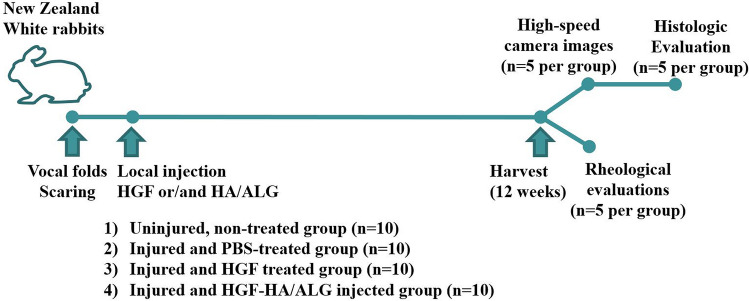
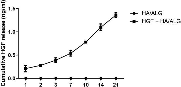
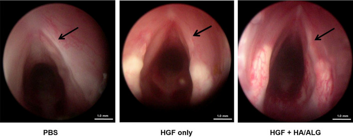
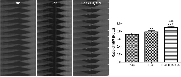
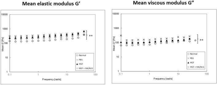
Methods: HGF was loaded into HA/ALG composite hydrogel (HGF-HA/ALG) and its in vitro release profile was evaluated. In addition, HGF-HA/ALG was injected into the VFs of rabbits immediately after direct injury and HGF or PBS was injected in the same manner into control groups. Macroscopic features were observed by endoscopy at 3 months post-injury. Functional analyses including mucosal waves of VFs and viscoelastic properties were performed by kymography following high-speed digital imaging and rheometer. Histopathological and immunohistochemical evaluations were also conducted on VFs.

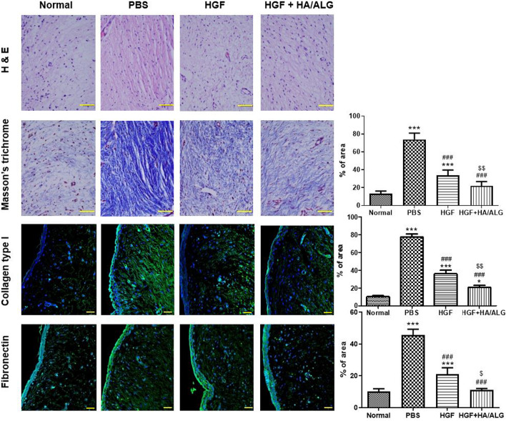
Results: HGF release from HGF-HA/ALG was sustained for up to 3 weeks. Rabbits treated with HGF-HA/ALG showed improved mucosal vibrations and VF viscoelastic properties as compared with the PBS and HGF controls. Histopathological staining revealed HGF-HA/ALG treated VFs showed less fibrosis than PBS and HGF controls, and immunohistochemical analysis demonstrated amounts of type I collagen and fibronectin were lower in HGF-HA/ALG treated animals than in PBS and HGF controls at 3 months post-injury.

Conclusion: HGF containing HA/ALG hydrogel enhanced healing in our rabbit model of VF injury.

Keywords: Alginate; Hepatocyte growth factor; Hyaluronic acid; Vocal fold; Wound healing.


Conflict of interest statement

The authors have no potential conflict of interest to declare.

Figures

Fig. 1
Fig. 1
Experimental flow chart of in vivo study design
Fig. 2
Fig. 2
Profile of hepatocyte growth factor (HGF) release from HA/ALG hydrogel. The amount of HGF released increased consistently over 21 days
Fig. 3
Fig. 3
Endoscopic examination of VFs at 3 months post-injury. Endoscopic examination of VFs at 3 months post-injury revealed prominent fibrotic scars in the PBS group, but reduced scar formation in the HGF and HGF–HA/ALG groups (the PBS group; injured and PBS treated, the HGF group; injured and HGF treated, HGF–HA/ALG group; injured and HGF–HA/ALG treated)
Fig. 4
Fig. 4
Micromorphological and immunohistochemical evaluations of ECM remodeling by HGF released from HGF–HA/ALG at 3 months post-injury. Scale bars represent 30 μm. Masson's trichrome (blue-stained collagen), collagen type I and fibronectin densities were quantified by summing numbers of pixels in positively stained areas using a software program. Results are presented as mean areas (%) ± SDs as determined by the Kruskal–Wallis test with Dunns’ post hoc test. *versus normal uninjured/non-treated controls, #versus injured/PBS treated animals, $versus injured/HGF treated animals, *p < 0.05, ***p < 0.001, ###p < 0.001, $p < 0.05 and $$p < 0.01. (n = 5 rabbits per group) (the normal control group; non-injured and non-treated, the PBS group; injured and PBS treated, the HGF group; injured and HGF treated, HGF–HA/ALG group; injured and HGF–HA/ALG treated)
Fig. 5
Fig. 5
Evaluation of mucosal wave oscillations after vocal fold injury. The amplitudes of mucosal waves (MWs) of VFs were evaluated by measuring the pixels of amplitudes of a mid-point of upper lips on high-speed digital images using image analysis software at 3 months post-injury. The amplitude ratios of MWs of treated VFs versus normal controls were calculated using Metamorph software. Normalized MW results are presented as mean ratios (right VFs to left VFs) ± SDs. Kruskal–Wallis test with Dunns’ post hoc test. *versus the PBS group, #versus the HGF group, **p < 0.01, ***p < 0.001 and ###p < 0.001 (n = 5 rabbits per group) (PBS group; injured and PBS treated, HGF group; injured and HGF treated, HGF–HA/ALG group; injured and HGF–HA/ALG treated)
Fig. 6
Fig. 6
Rheometric evaluations of viscoelasticity changes after vocal fold injury. Viscoelastic properties of regenerated VFs were measured at 3 months post-injury. Mean elastic moduli (G′) and viscous moduli (G″) are plotted on a log–log scale as a function of frequency. Rheological data were analyzed at all frequencies by two-way ANOVA with Bonferroni’s post hoc test (both p < 0.01) (n = 5 rabbits per group) (the normal control group; non-injured and non-treated, the PBS group; injured and PBS treated, the HGF group; injured and HGF treated, HGF–HA/ALG group; injured and HGF–HA/ALG treated)

References

    1. Choi JS, Lee S, Kim DY, Kim YM, Kim MS, Lim JY. Functional remodeling after vocal fold injury by small intestinal submucosa gel containing hepatocyte growth factor. Biomaterials. 2015;40:98–106. doi: 10.1016/j.biomaterials.2014.11.028. - DOI - PubMed
    1. Hansen JK, Thibeault SL. Current understanding and review of the literature: vocal fold scarring. J Voice. 2006;20:110–120. doi: 10.1016/j.jvoice.2004.12.005. - DOI - PubMed
    1. Woo SH, Kim RB, Choi SH, Lee SW, Won SJ. Prevalence of laryngeal disease in South Korea: data from the Korea National Health and Nutrition Examination Survey from 2008 to 2011. Yonsei Med J. 2014;55:499–507. doi: 10.3349/ymj.2014.55.2.499. - DOI - PMC - PubMed
    1. Long JL. Tissue engineering for treatment of vocal fold scar. Curr Opin Otolaryngol Head Neck Surg. 2010;18:521–525. doi: 10.1097/MOO.0b013e32833febf2. - DOI - PubMed
    1. Hirano S, Bless DM, Rousseau B, Welham N, Montequin D, Chan RW, et al. Prevention of vocal fold scarring by topical injection of hepatocyte growth factor in a rabbit model. Laryngoscope. 2004;114:548–556. doi: 10.1097/00005537-200403000-00030. - DOI - PubMed

Publication types

LinkOut - more resources