. 2021 Jul;17(7):1753-1767.
doi: 10.1080/15548627.2020.1783822. Epub 2020 Jul 17.

Autophagy core protein ATG5 is required for elongating spermatid development, sperm individualization and normal fertility in male mice

Affiliations

Autophagy core protein ATG5 is required for elongating spermatid development, sperm individualization and normal fertility in male mice

Qian Huang et al. Autophagy. 2021 Jul.

Abstract

Spermiogenesis is the longest phase of spermatogenesis, with dramatic morphological changes and a final step of spermiation, which involves protein degradation and the removal of excess cytoplasm; therefore, we hypothesized that macroautophagy/autophagy might be involved in the process. To test this hypothesis, we examined the function of ATG5, a core autophagy protein in male germ cell development. Floxed Atg5 and Stra8- iCre mice were crossed to conditionally inactivate Atg5 in male germ cells. In Atg5flox/flox; Stra8- iCre mutant mice, testicular expression of the autophagosome marker LC3A/B-II was significantly reduced, and expression of autophagy receptor SQSTM1/p62 was significantly increased, indicating a decrease in testicular autophagy activity. The fertility of mutant mice was dramatically reduced with about 70% being infertile. Sperm counts and motility were also significantly reduced compared to controls. Histological examination of the mutant testes revealed numerous, large residual bodies in the lumen of stages after their normal resorption within the seminiferous epithelium. The cauda epididymal lumen was filled with sloughed germ cells, large cytoplasmic bodies, and spermatozoa with disorganized heads and tails. Examination of cauda epididymal sperm by electron microscopy revealed misshapen sperm heads, a discontinuous accessory structure in the mid-piece and abnormal acrosome formation and loss of sperm individualization. Immunofluorescence staining of epididymal sperm showed abnormal mitochondria and acrosome distribution in the mutant mice. ATG5 was shown to induce autophagy by mediating multiple signals to maintain normal developmental processes. Our study demonstrated ATG5 is essential for male fertility and is involved in various aspects of spermiogenesis.Abbreviations: AKAP4: a-kinase anchoring protein 4; ATG5: autophagy-related 5; ATG7: autophagy-related 7; ATG10: autophagy-related 10; ATG12: autophagy-related 12; cKO: conditional knockout; DDX4: DEAD-box helicase 4; MAP1LC3/LC3/tg8: microtubule-associated protein 1 light chain 3; PBS: phosphate-buffered saline; PIWIL2/MILI: piwi like RNA-mediated gene silencing 2; RT-PCR: reverse transcription-polymerase chain reaction; SQSTM1/p62: sequestosome 1; TBC: tubulobulbar complexes; WT: wild type.

Keywords: ATG5; Acrosome; autophagy; individualization; male germ cells; male reproduction; mitochondria; spermiogenesis.


Conflict of interest statement

There is no conflict of interest that could be perceived as prejudicing the impartiality of the research reported.

Figures

Figure 1.
Figure 1.
ATG5 is abundant in mouse testis and is present in the cytoplasm of spermatocytes and round spermatids. (A) Analysis of tissue distribution of mouse ATG5 protein by western blot. ATG5 protein was abundant in the testis. It was also detected in the liver, kidney and spleen in our experiment (upper panel). GAPDH was analyzed as a loading control (bottom panel). (B) Examination of ATG5 localization in isolated male germ cells by immunofluorescence staining. Isolated germ cells were stained with the ATG5 antibody (red). The ATG5 signal was present as cytoplasmic vesicles in spermatocytes and round spermatids
Figure 2.
Figure 2.
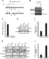
Generation of Atg5 conditional knockout (cKO) mice and the decreased autophagy activity in the testis of the Atg5 cKO mice. (A) Schematic representation of the mouse Atg5 gene structure, strategy to target exon 3 and locations of primers used in the study. (B) Representative RT-PCR results using primers that amplify exons 2 to 4 of Atg5 cDNA. a DNA band with smaller size was detected in the mutant mice, indicating that exon 3 was spliced out. (C) Real-time PCR results showed that Atg5 mRNA expression level was significantly reduced in the knockout mice. (D) Representative western blot result showing testicular ATG5 was absent in the atg5 cKO mice. (E) Expression of LC3A/B and SQSTM1/p62 as examined by western blot in the control and conditional atg5 knockout mice (n = 3). The level of 14 kDa LC3A/B-II, an indicator of active autophagy, was hardly detected in the mutant mice; SQSTM1, an autophagy substrate, was increased, indicating decreased autophagy activity in the testis of atg5 cKO mice. (F) Quantification of the relative LC3A/B-I (i) and SQSTM1 (ii) protein expression. Error bars represent the standard deviation (n = 3). (*), p < 0.05
Figure 3.
Figure 3.
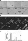
Abnormal sperm morphologies in the atg5 cKO mice. (A) Representative epididymal sperm of control (i) and atg5 cKO mice (ii–viii) examined by DIC microscopy. Sperm in the control mice showed normal morphology (i). Multiple abnormalities were observed in the atg5 cKO mice, including distorted (arrow) and round heads (arrowhead) (ii), abnormal flagella (ii–iv), double heads (v), and sperm bundle (vi–viii). (B) Representative epididymal sperm of control (i) and atg5 cKO mice (ii–Viii) examined by scanning electron microscopy. As observed under DIC microscope, well-developed head (H) and long and smooth flagella (F) were present in the control mice (i), the atg5 cKO sperm had abnormally developed sperm heads (arrowheads in ii, iii, iv, vi, vii), discontinuous accessory structure in the middle piece were also observed in some sperm (arrowheads, iii, iv, vii, viii); some sperm had distorted structure in the middle piece (v), short tail (vi), sperm bundle was also seen (viii). (C) Quantification of the percentage of different types of abnormal sperm in the control and atg5 cKO mice
Figure 4.
Figure 4.
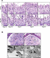
Histology of testis and epididymis from the control and atg5 cKO mice. (A) Testis histology from control (WT) and atg5 cKO mice showing sections of the seminiferous epithelium. Images correspond to the respective stages of spermatogenesis from control testis (top row) and the atg5 cKO testis (bottom row). The numbers represent the various steps of round and elongated spermatids. M1 labels the pachytene spermatocytes in meiotic division in stage XII. In all stages of the atg5 cKO testis, spermatogonia, spermatocytes and round spermatids appear normal compared to controls. However, the cytoplasm of elongating spermatids begins to appear abnormal in step 14–16, with the sloughing of cells into the lumen along with round bodies of cytoplasm and the fusion of some elongating cells into giant bodies. Scale bar = 20 μm for all photos. (I) WT stage II–III showing bundles of elongating step 14 and round step 2–3 spermatids. (ii) WT stage V–VI, with bundles of step 15 spermatids resting above the round spermatids. (iii) WT stage IX. Step 9 spermatid nuclei have begun to show the angling of the nucleus at the start of elongation. Step 16 spermatids have matured and released into the lumen as sperm. Residual bodies have been reabsorbed rapidly by the Sertoli cells in this stage and are rarely seen in stages IX–X. (iv) WT stage XI, with an abundance of step 11 elongating spermatids lining the lumen. (V) WT stage XII. Bundles of step 12 spermatids sit above the dividing pachytene spermatocytes that exhibit meiotic division (M1). (vi) atg5 cKO stage II–III. Step 2–3 round spermatids appear normal; however, the cytoplasm of elongating step 14 spermatids appear abnormal, with the sloughing of cells into the lumen along with round bodies of cytoplasm (Cy). The elongating spermatid bundles have fewer numbers of cells. (vii) atg5 cKO stage V–VI. The elongating step 15 spermatids have abnormal heads and there is fusion of some elongating cells into giant bodies. (viii) atg5 cKO stage IX, with step 16 spermatids show retention in the epithelium, rather than being released by spermiation. Abnormally large bodies of fused residual bodies (Rb) are seen sloughing into the lumen and subsequently appear in the epididymis. In the KO testis, the fused residual bodies were often found in stages XI–XII, along with the retained step 16 spermatids. (ix) atg5 cKO stage XI, showing residual bodies (Rb) at the lumen, but also with attached step 16 spermatids being phagocytosed by the epithelium and near the basement membrane. (X) atg5 cKO stage XI, with large residual bodies (Rb) near the lumen along with step 16 spermatids being retained. (xi) atg5 cKO stage XII. Abnormal step 12 spermatids are lacking bundle formation, indicating that there has been the loss of elongating spermatids due to sloughing and phagocytosis. Step 16 spermatid heads are seen near the basement membrane, indicating phagocytosis. (xii) atg5 cKO stage XII. Meiotic division (M1) of the pachytene spermatocytes appears to be normal, but the elongating spermatids show abnormalities and fewer numbers near the lumen. Step 9 spermatids are also abnormally present near the lumen. (B) Significant number of residual bodies were present in the epididymis and seminiferous tubule lumen of the atg5 cKO mice. (i) Control cauda epididymis showing the epithelium (Ep) lining the lumen that is filled with normal sperm aligned with their heads (Hd) and tails (T). (ii) atg5 cKO cauda epididymis showing a lumen filled with numerous, large cytoplasmic bodies (Cy), sloughed germ cells (Gc) and spermatozoa with heads (Hd) and tails (T) that are less aligned than those in the control. Ep, epithelium. (iii) TEM of lumen area of seminiferous tubules of control mice. Normally developing spermatids in a control mouse with well-developed heads (arrows). (iv) TEM of lumen area of seminiferous tubules of atg5 cKO mice. Residual bodies (arrows) were present, and abnormally formed chromatin (arrowheads) was throughout. The dashed arrows may represent lipid droplets and RNAs (leftover ribosomes)
Figure 5.
Figure 5.
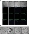
Abnormal acrosome biogenesis in the atg5 cKO mice. (A) Immunofluorescence staining was performed on cauda epididymal sperm from control (left panel) and atg5 cKO mice (right three panels). Peanut-lectin was used as a marker for acrosome. The images show the abnormal acrosome formation in the atg5 cKO mice. (B) Examination of spermatid acrosome in the testis seminiferous tubules of control (i, ii) and atg5 cKO mice (iii, iv) by electronic microscopy. In the control mouse, normal acrosome development can be seen in the round and elongating spermatids (arrows in i and ii). However, in the atg5 cKO mice, abnormal acrosome development was present (dashed arrows in iii and iv)
Figure 6.
Figure 6.
Abnormal sperm mitochondrial sheath formation in the atg5 cKO mice. (A) Representative images of mitochondria staining of cauda epididymal sperm from control and atg5 cKO mice. Normal sperm mitochondria staining (i) can be seen in the control mouse. Discontinuous (arrowhead, ii) and bundle (arrows, iii, iv) mitochondria signal was seen in the atg5 cKO mouse. (B) Representative TEM images of epididymal sperm from control and atg5 cKO mice. a control mouse showed the separate axoneme formed around with connected mitochondria (arrowheads and the insert, i). In the atg5 cKO mouse, several axonemes were included in one member (arrows, ii, iii) and some of them were surrounded with disconnected mitochondria (arrowheads, iii, iv)
Figure 7.
Figure 7.
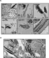
Defective sperm chromatin structure and disrupted individualization in the atg5 cKO mice as revealed by TEM study. (A) Epididymal sperm. (i) a representative image showing a large number of sperm collected from cauda epididymis. Normal chromatin structure was present (insert) in a control mouse. (ii, iii, iv) The arrows point to the abnormally formed chromatin of epididymal sperm from the atg5 cKO mouse; (iv, v, vi) multiple sperm were wrapped in one cell membrane (arrowheads). (B) Seminiferous tubules. (i) a representative image showing normally separated elongating spermatids in the seminiferous tubules of a control mouse. The arrow points to the cell membrane; (ii) multiple elongating spermatids in the seminiferous tubule were wrapped in one cell membrane in the atg5 cKO mouse, indicating failure of individualization. The arrowheads point to the elongating spermatids sharing the same cell membrane

References

    1. Feng Y, He D, Yao Z, et al. The machinery of macroautophagy. Cell Res. 2014;24:24. - PMC - PubMed
    1. Yoshii SR, Mizushima N.. Monitoring and measuring autophagy. Int J Mol Sci. 2017;18:1865. - PMC - PubMed
    1. Mizushima N, Levine B. Autophagy in mammalian development and differentiation. Nat Cell Biol. 2010;12:823. - PMC - PubMed
    1. Noda NN, Inagaki F. Mechanisms of autophagy. Annu Rev Biophys. 2015;44:101–122. - PubMed
    1. Nakatogawa H. Two ubiquitin-like conjugation systems that mediate membrane formation during autophagy. Essays Biochem. 2013;55:39–50. - PubMed

Publication types

Substances