

Metastatic Calcinosis of Gastric Mucosa
- PMID: 32677845
- PMCID: PMC7370329
- DOI: 10.1177/2324709620940482
Metastatic Calcinosis of Gastric Mucosa
Abstract
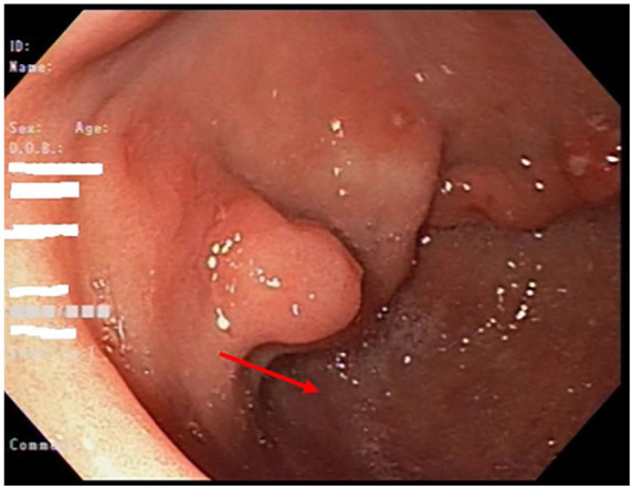
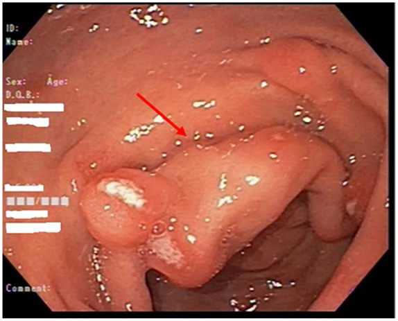
Calcinosis cutis refers to the deposition of calcium salts in the cutaneous and subcutaneous tissue and is frequently associated with inflammation. Gastric calcinosis can be classified into metastatic, dystrophic, and idiopathic; metastatic calcinosis is the most common type. In metastatic calcification, calcium salts are deposited in normal soft tissues in the setting of altered metabolism of serum calcium and phosphorus and is a rare and serious complication of chronic renal failure. The important factors contributing to the development of metastatic calcinosis are hypercalcemia, hyperphosphatemia, and an elevated calcium-phosphate product. The most striking feature of this diagnosis is the calcification around the large joints. While it mostly involves dermis of small and medium-sized vessels, it can rarely affect the mucosal layers of the gastrointestinal (GI) tract. Calcinosis presents as a marker for the presence of calcifications in other organs, such as heart or lung, which can be life-threatening. Patients rarely present with clinical symptoms of GI upset, dyspepsia, or epigastric pain that are attributed to calcinosis. If patients present with GI symptoms, infectious causes remain to be higher on the differential. We present a case of incidental finding of gastric mucosal calcinosis during the workup and treatment of dysphagia.
Keywords: chronic kidney disease; hypercalcemia; hyperphosphatemia; metastatic calcinosis cutis; metastatic gastric calcinosis.
Conflict of interest statement
Figures


References
-
- Gorospe M, Fadare O. Gastric mucosal calcinosis: clinicopathologic considerations. Adv Anat Pathol. 2007;14:224-228. - PubMed
-
- Hsieh TH, McCullough A, Aqel B. Gastric mucosal calcinosis. Gastrointest Endosc. 2011;73:1282-1283. - PubMed
-
- Reiter N, El-Shabrawi L, Leinweber B, Berghold A, Aberer E. Calcinosis cuits: part I. Diagnostic pathway. J Am Acad Dermatol. 2011;65:1-12. - PubMed
-
- Mulligan RM. Metastatic calcification. Arch Pathol (Chic). 1947;43:177-230. - PubMed
Publication types
MeSH terms
Substances
LinkOut - more resources
Full Text Sources
Medical
