. 2020 Jul 16;17(1):107.
doi: 10.1186/s12985-020-01357-3.

Influenza A virus NS1 optimises virus infectivity by enhancing genome packaging in a dsRNA-binding dependent manner

Affiliations

Influenza A virus NS1 optimises virus infectivity by enhancing genome packaging in a dsRNA-binding dependent manner

Tim Wai Sha et al. Virol J. .

Abstract

Background: The non-structural protein 1 (NS1) of influenza A virus (IAV) is a key player in inhibiting antiviral response in host cells, thereby facilitating its replication. However, other roles of NS1, which are independent of antagonising host cells' antiviral response, are less characterised.

Methods: To investigate these unidentified roles, we used a recombinant virus, which lacks NS1 expression, and observed its phenotypes during the infection of antiviral defective cells (RIG-I KO cells) in the presence or absence of exogeneous NS1. Moreover, we used virus-like particle (VLP) production system to further support our findings.

Results: Our experiments demonstrated that IAV deficient in NS1 replicates less efficiently than wild-type IAV in RIG-I KO cells and this replication defect was complemented by ectopic expression of NS1. As suggested previously, NS1 is incorporated in the virion and participates in the regulation of viral transcription and translation. Using the VLP production system, in which minigenome transcription or viral protein production was unaffected by NS1, we demonstrated that NS1 facilitates viral genome packaging into VLP, leading to efficient minigenome transfer by VLP. Furthermore, the incorporation of NS1 and the minigenome into VLP were impaired by introducing a point mutation (R38A) in the double stranded RNA-binding domain of NS1.

Conclusion: These results suggest a novel function of NS1 in improving genome packaging in a dsRNA binding-dependent manner. Taken together, NS1 acts as an essential pro-viral regulator, not only by antagonizing host immunity but also by facilitating viral replication and genome packaging.

Keywords: Genome packaging; Influenza a virus; Innate immunity; Non-structural protein 1; Virus-like production system; dsRNA binding domain.


Conflict of interest statement

The authors declare that they have no competing interests.

Figures

Fig. 1
Fig. 1
IAV replication in WT and RIG-I KO cells. a 293 T RIG-I KO cells were mock treated or infected with indicated viruses at MOI = 0.1. Twenty-four hours after infection, extracted total RNA was reverse-transcribed using random primers followed by qPCR with primers targeting IFNB1. WT 293 T (b) and RIG-I KO cells (c) were infected with PR8-WT or PR8-delNS1 at MOI 0.01. The virus yield was measured at 48 h post-infection by plaque assay using MDCK cells. The Student’s t test was used for statistical analysis (**P < 0.01, *P < 0.05). The data shown are the mean ± standard deviation from at least two independent experiments (n = 2 in (a), n = 3 in (b) & (c))
Fig. 2
Fig. 2
Effects of NS1 on virus yield. a 293 T RIG-I KO cells were infected with PR8-delNS1 at MOI 0.01 for 1 h and immediately mock transfected or transfected with empty plasmid control (E.C.) or those encoding GFP or NS1 protein. Virus yields were measured 48 h post-infection by plaque assay using MDCK cells. The data shown are the mean ± standard deviation from at least three independent experiments (n = 3). The Student’s t test was used for statistical analysis (n.s. not significant, **P < 0.01). b 293 T RIG-I KO cells were infected as in (a). Cells and culture supernatant were collected to prepare cell lysate and purified virion, respectively. Equal volumes of purified virus were subjected to immunoblot analysis using anti-NP, anti-M1, anti-HA and anti-lamin B antibodies. c 293 T RIG-I KO cells were infected with PR8-delNS1 at MOI 0.1 for 1 h and immediately transfected with NS1 expression plasmid or empty control plasmid. Cells were stained by fluorophore probe targeting segment 5 vRNA (vRNA, Red), anti- NS1 (green), anti-M1 (white), anti-HA (white) and DAPI (blue) 24 h after infection and transfection, and imaged by confocal microscopy. Scale bar, 10 μm. d Supernatant collected from (a) and virus quantity (HAU) was determined by HA assay using 0.5% chicken RBC. e vRNA was extracted from equal HAU from (a) and quantified by RT-qPCR. Levels of viral genome of virus containing supernatant obtained from E.C. transfected cells were set to 1. The Student’s t test was used for statistical analysis (*P < 0.05, **P < 0.01). The data shown are the mean ± standard deviation from at least three independent experiments (n = 3). f Cells in (a) were subjected to extraction for total RNA and vRNA was quantified by qPCR. Levels of viral genome obtained from E.C. transfected cells were set to 1. The Student’s t test was used for statistical analysis (n.s. not significant, *P < 0.05, **P < 0.01). The data shown are the mean ± standard deviation from at least three independent experiments (n = 3)
Fig. 3
Fig. 3
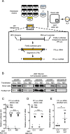
Effects of NS1 on viral replication in the VLP system. a 293 T RIG-I KO cells were co-transfected with eleven expression plasmids as described and a minigenome expression construct. Minigenome expression is regulated by RNA pol I as illustrated. b Twenty-four hours after transfection, cells and VLP released in the culture medium were harvested. VLP was further purified (Materials and Methods). Proteins in donor cells and purified VLP were detected by immunoblotting. c Luciferase activity in the donor cells was measured. Firefly luciferase activity was normalised by Renilla luciferase activity. Luciferase activity without PB1 expression was set to 1. The Student’s t test was used for statistical analysis (n.s. not significant). The data shown are the mean ± standard deviation from at least three independent experiments (n = 3). d Total RNA was extracted from the donor cells and minigenome was quantified by strand specific RT-qPCR. Levels of firefly luciferase vRNA obtained from cells without PB1 expression were set to 1. The Student’s t test was used for statistical analysis (n.s. not significant). The data shown are the mean ± standard deviation from at least three independent experiments (n = 3). e vRNA in the purified VLP was quantified by strand specific RT-qPCR. Levels of Firefly luciferase vRNA obtained from E.C. transfected cells were set to 1. The Student’s t test was used for statistical analysis (*P < 0.05). The data shown are the mean ± standard deviation from at least three independent experiments (n = 3)
Fig. 4
Fig. 4
Effects of NS1 on gene transduction and infection by VLP in the recipient cells. a Schematic representation of VLP production and minigenome gene transduction and infection in the recipient cells. b VLP produced in the culture supernatant was transferred to the recipient cells derived either from WT 293 T or 293 T RIG-I KO cells. Luciferase activity was measured after 8 h. The Student’s t test was used for statistical analysis (*P < 0.05, **P < 0.01). The data shown are the mean ± standard deviation from at least three independent experiments (n = 3). c VLP-treated recipient cells (293 T RIG-I KO) were stained with anti-luciferase antibody (red) and DAPI (blue), and examined by confocal microscopy (left). Scale bar, 10 μm. Cells were quantified for luciferase expression (right). The Student’s t test was used for statistical analysis (*P < 0.05). The data shown are the mean ± standard deviation from at least three independent experiments (n = 3)
Fig. 5
Fig. 5
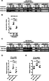
Involvement of RBD of NS1 in viral replication. a 293 T RIG-I KO cells were infected with PR8 delNS1 at MOI 0.01 and transfected with empty vector (E.C.), NS1 WT or NS1 R38A as described in Fig. 2a. Cell lysate and purified virion were analysed by immunoblotting. b Viruses released in the culture supernatant were quantified by plaque assay. The Student’s t test was used for statistical analysis (n.s. not significant, **P < 0.01). The data shown are the mean ± standard deviation from at least three independent experiments (n = 3). c 293 T RIG-I KO cells were used to produce VLP as described in Fig. 3a in the absence of NS1 or NS1 WT or NS1 R38A. Proteins in the donor cells or purified VLP were examined by immunoblotting. d vRNA extracted from VLP was quantified by RT-qPCR. Levels of Firefly luciferase vRNA obtained from E.C. transfected cells were set to 1. The Student’s t test was used for statistical analysis (n.s. not significant, *P < 0.05). The data shown are the mean ± standard deviation from at least three independent experiments (n = 3). e VLP was transferred to the recipient cells (293 T RIG-I KO, Fig. 4a) and luciferase activity was measured. The Student’s t test was used for statistical analysis (n.s. not significant, **P < 0.01). The data shown are the mean ± standard deviation from at least three independent experiments (n = 3)

Similar articles

Cited by

References

    1. Stasakova J, Ferko B, Kittel C, Sereinig S, Romanova J, Katinger H, Egorov A. Influenza a mutant viruses with altered NS1 protein function provoke caspase-1 activation in primary human macrophages, resulting in fast apoptosis and release of high levels of interleukins 1beta and 18. J Gen Virol. 2005;86(Pt 1):185–195. doi: 10.1099/vir.0.80422-0. - DOI - PubMed
    1. Wang X, Li M, Zheng H, Muster T, Palese P, Beg AA, Garcia-Sastre A. Influenza a virus NS1 protein prevents activation of NF-kappaB and induction of alpha/beta interferon. J Virol. 2000;74(24):11566–11573. doi: 10.1128/JVI.74.24.11566-11573.2000. - DOI - PMC - PubMed
    1. Moriyama M, Chen IY, Kawaguchi A, Koshiba T, Nagata K, Takeyama H, Hasegawa H, Ichinohe T. The RNA- and TRIM25-binding domains of influenza virus NS1 protein are essential for suppression of NLRP3 Inflammasome-mediated interleukin-1beta secretion. J Virol. 2016;90(8):4105–4114. doi: 10.1128/JVI.00120-16. - DOI - PMC - PubMed
    1. Chung WC, Kang HR, Yoon H, Kang SJ, Ting JP, Song MJ. Influenza a virus NS1 protein inhibits the NLRP3 Inflammasome. PLoS One. 2015;10(5):e0126456. doi: 10.1371/journal.pone.0126456. - DOI - PMC - PubMed
    1. Talon J, Horvath CM, Polley R, Basler CF, Muster T, Palese P, Garcia-Sastre A. Activation of interferon regulatory factor 3 is inhibited by the influenza a virus NS1 protein. J Virol. 2000;74(17):7989–7996. doi: 10.1128/JVI.74.17.7989-7996.2000. - DOI - PMC - PubMed

Publication types