Review
. 2020 Jul 16;6(1):58.
doi: 10.1038/s41572-020-0188-7.

Inherited cardiac arrhythmias

Affiliations
Review

Inherited cardiac arrhythmias

Peter J Schwartz et al. Nat Rev Dis Primers. .

Abstract

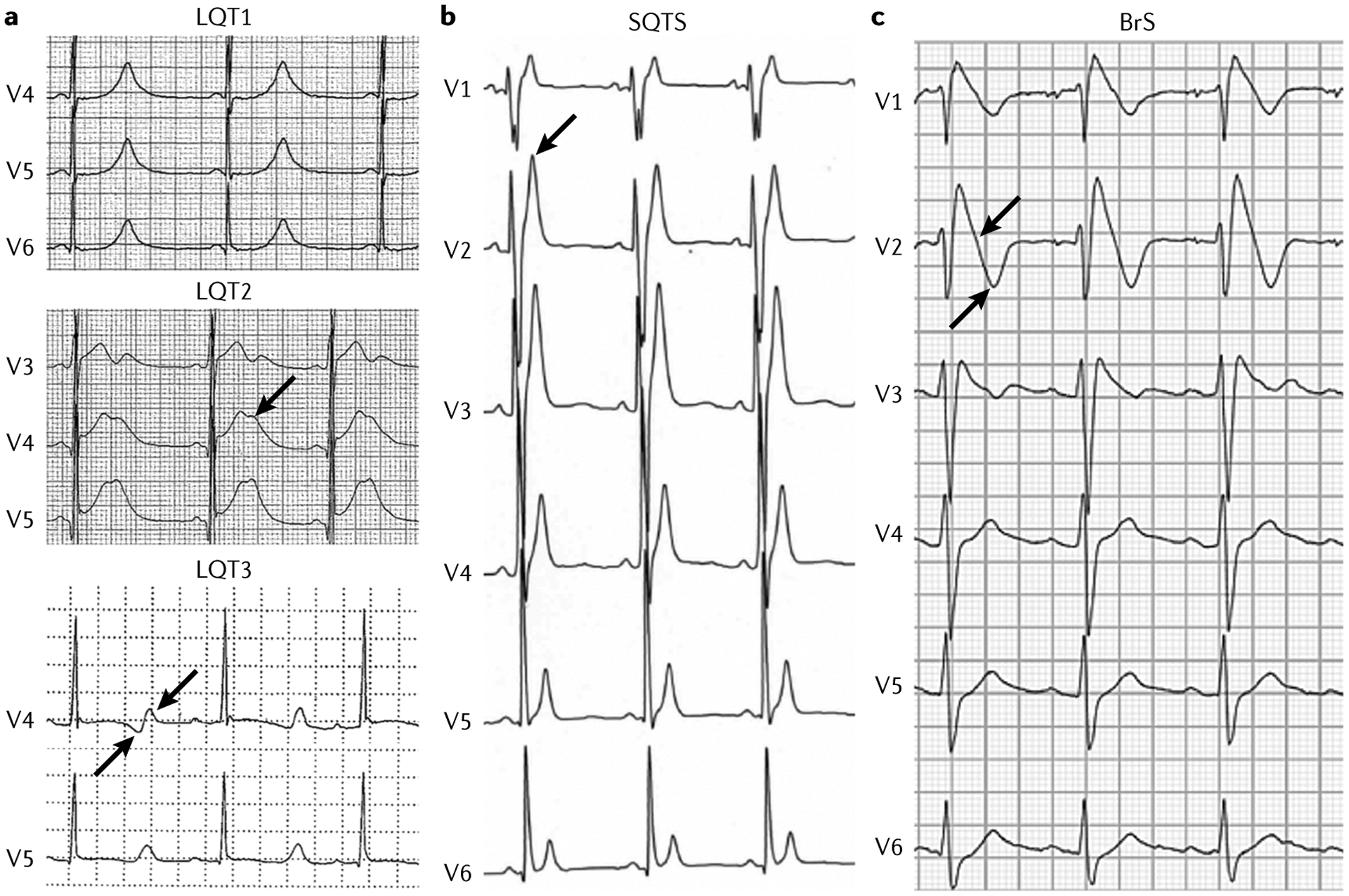
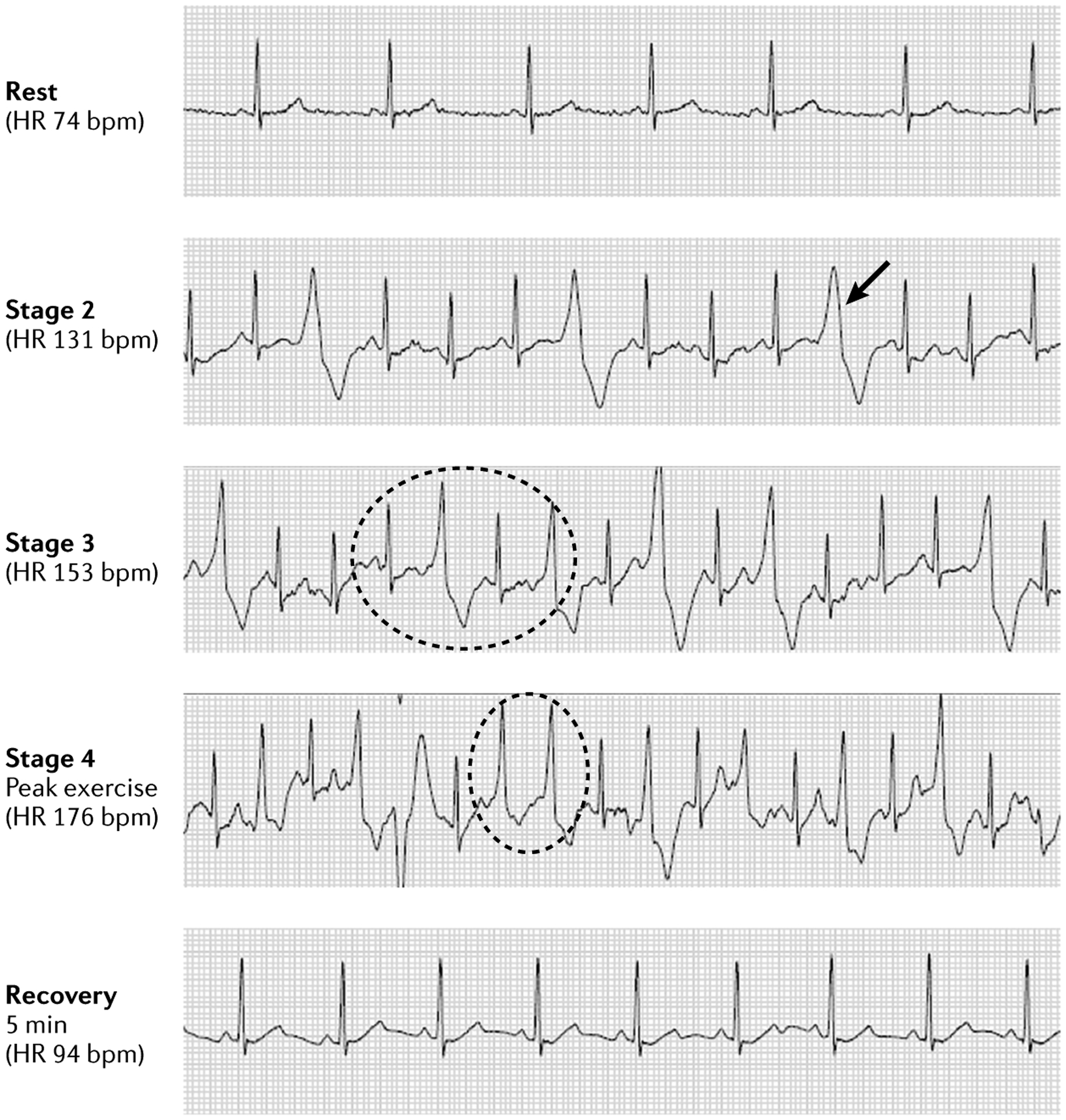
The main inherited cardiac arrhythmias are long QT syndrome, short QT syndrome, catecholaminergic polymorphic ventricular tachycardia and Brugada syndrome. These rare diseases are often the underlying cause of sudden cardiac death in young individuals and result from mutations in several genes encoding ion channels or proteins involved in their regulation. The genetic defects lead to alterations in the ionic currents that determine the morphology and duration of the cardiac action potential, and individuals with these disorders often present with syncope or a life-threatening arrhythmic episode. The diagnosis is based on clinical presentation and history, the characteristics of the electrocardiographic recording at rest and during exercise and genetic analyses. Management relies on pharmacological therapy, mostly β-adrenergic receptor blockers (specifically, propranolol and nadolol) and sodium and transient outward current blockers (such as quinidine), or surgical interventions, including left cardiac sympathetic denervation and implantation of a cardioverter-defibrillator. All these arrhythmias are potentially life-threatening and have substantial negative effects on the quality of life of patients. Future research should focus on the identification of genes associated with the diseases and other risk factors, improved risk stratification and, in particular for Brugada syndrome, effective therapies.


Conflict of interest statement

Competing interests

M.J.A. is a consultant for Audentes Therapeutics, Boston Scientific, Gilead Sciences, Invitae, Medtronic, MyoKardia and St. Jude Medical, and holds equity/royalties of AliveCor, Blue Ox Health and StemoniX. C.A. is a consultant for Novartis Institutes for BioMedical Research, Inc. and Trevena, Inc. All other authors declare no competing interests.

Figures

Fig. 1 |
Fig. 1 |. Genes and proteins involved in the pathogenesis of inherited cardiac arrhythmias.
The figure shows the transmembrane ionic channels that are responsible for the potassium (IKs, IK1 and IKr), calcium (ICa) and sodium (INa) currents that contribute to the cardiac action potential. Proteins of the sarcoplasmic reticulum that are involved in calcium handling (encoded by CASQ2, RYR2, CALM1, CALM2, CALM3 and TRDN) are also shown. Other proteins that can be mutant in inherited cardiac arrhythmias are encoded by ANK2 and TECRL. The genes that encode all these proteins or their subunits are coloured according to the disease with which they have been associated. BrS, Brugada syndrome; CPVT, catecholaminergic polymorphic ventricular tachycardia; LQTS, long QT syndrome; SQTS, short QT syndrome.
Fig. 2 |
Fig. 2 |. Variability in baseline QTc.
Distribution of the duration of heart rate corrected QT interval (QTc) in individuals who carry the same KCNQ1 mutation and who belong to a LQT1 South African founder population. The vertical dashed line represents the upper limit of normal values for men (440 ms). If the QTc prolongation were determined only by the A341V mutation, the QTc values should be uniformly prolonged with modest variability. The large spectrum of QTc values points to the presence of additional variants affecting ventricular repolarization. Data from REF..
Fig. 3 |
Fig. 3 |. Ventricular action potential and ionic currents.
a | Schematic showing a normal electrocardiogram trace and the corresponding phases of the ventricular action potential (0, 1, 2, 3 and 4) that determine the shape of the trace. b | The major transmembrane ionic currents that generate the ventricular action potential. Inward currents that contribute to depolarization are oriented downwards, and outward currents that contribute to repolarization are oriented upwards; the shapes of the currents indicate their relative intensity. Ito, transient outward potassium current.
Fig. 4 |
Fig. 4 |. Spatial dispersion of repolarization.
a | A prominent transient outward current (Ito) is responsible for phase 1 of the action potential (AP), giving rise to a prominent AP notch in the epicardium but not in the endocardium, where this current is relatively small. b | The presence of a prominent notch in the epicardium but not the endocardium gives rise to a transmembrane voltage gradient. Heterogeneous transmural distribution of the Ito-mediated AP notch is responsible for the inscription of the J wave on the electrocardiogram (ECG). Part a adapted from REF., Liu, D. W., Gintant, G. A. & Antzelevitch, C. Ionic bases for electrophysiological distinctions among epicardial, midmyocardial, and endocardial myocytes from the free wall of the canine left ventricle. Circ. Res. 72(3), 671–687 (https://www.ahajournals.org/journal/res). Part b adapted from REF., Yan G. X. & Antzelevitch C. Cellular basis for the electrocardiographic J wave. Circulation 93(2), 372–379 (https://www.ahajournals.org/journal/circ).
Fig. 5 |
Fig. 5 |. Examples of ECG traces.
a | Example of electrocardiogram (ECG) traces of patients with long QT syndrome (LQTS). In LQTS type 1 (LQT1), the QT interval is extremely prolonged, with a tall peaked T wave. In LQT2, a very prolonged QT interval is present, with a clear and typical notch on the T wave (arrow). In LQT3, a very prolonged QT interval is followed by a late-onset diphasic (arrows) T wave. b | In a patient with short QT syndrome (SQTS), the QT interval is extremely short, with a tall peaked T wave (arrow). c | A patient with Brugada syndrome (BrS) has an ECG trace with a typical type 1 pattern. There is a coved-type ST segment (arrow) in the right precordial leads followed by a terminal negative T wave (arrow).
Fig. 6 |
Fig. 6 |. Catecholaminergic polymorphic ventricular tachycardia exercise test.
Exercise stress test in a patient with catecholaminergic polymorphic ventricular tachycardia as confirmed by genetic analysis. Sinus rhythm was normal at rest, with ectopy increasing with exercise. The patient demonstrated single premature ventricular contractions (PVCs) (arrow) with increasing frequency as the heart rate (HR) progressively increased, transitioning to PVCs in bigeminy (dotted circle in stage 3) with couplets (dotted circle in stage 4) and triplets at peak exercise. Ectopy disappears after 5 min of recovery, and normal sinus rhythm is restored. bpm, beats per minute.

References

    1. Meyer L et al. Incidence, causes, and survival trends from cardiovascular-related sudden cardiac arrest in children and young adults 0 to 35 years of age: a 30-year review. Circulation. 126, 1363–1372 (2012). - PubMed
    1. Winkel BG et al. Nationwide study of sudden cardiac death in persons aged 1–35 years. Eur. Heart J 32, 983–990 (2011). - PubMed
    1. Bardai A et al. Incidence, causes, and outcomes of out-of-hospital cardiac arrest in children. A comprehensive, prospective, population-based study in the Netherlands. J. Am. Coll. Cardiol 57, 1822–1828 (2011). - PubMed
    1. Bagnall RD et al. A prospective study of sudden cardiac death among children and young adults. N. Engl. J. Med 374, 2441–2452 (2016). - PubMed
    1. Kong MH et al. Systematic review of the incidence of sudden cardiac death in the United States. J. Am. Coll. Cardiol 57, 794–801 (2011). - PMC - PubMed

Publication types

MeSH terms