. 2020 Jul 16;10(1):11766.
doi: 10.1038/s41598-020-68685-w.

The novel features of Plantago ovata seed mucilage accumulation, storage and release

Affiliations

The novel features of Plantago ovata seed mucilage accumulation, storage and release

Jana L Phan et al. Sci Rep. .

Abstract

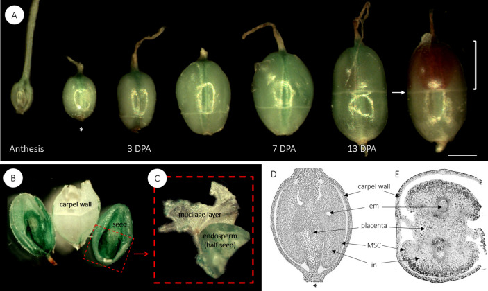
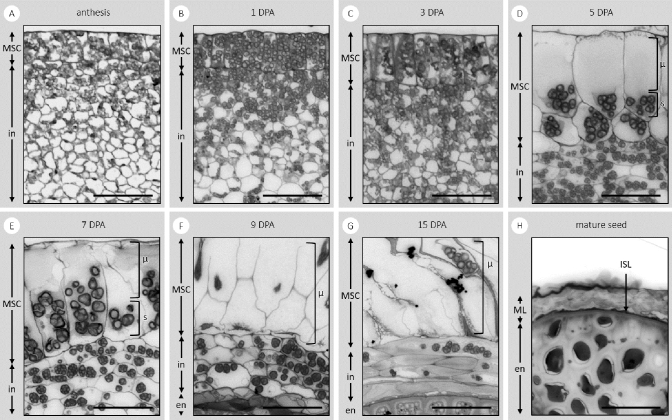
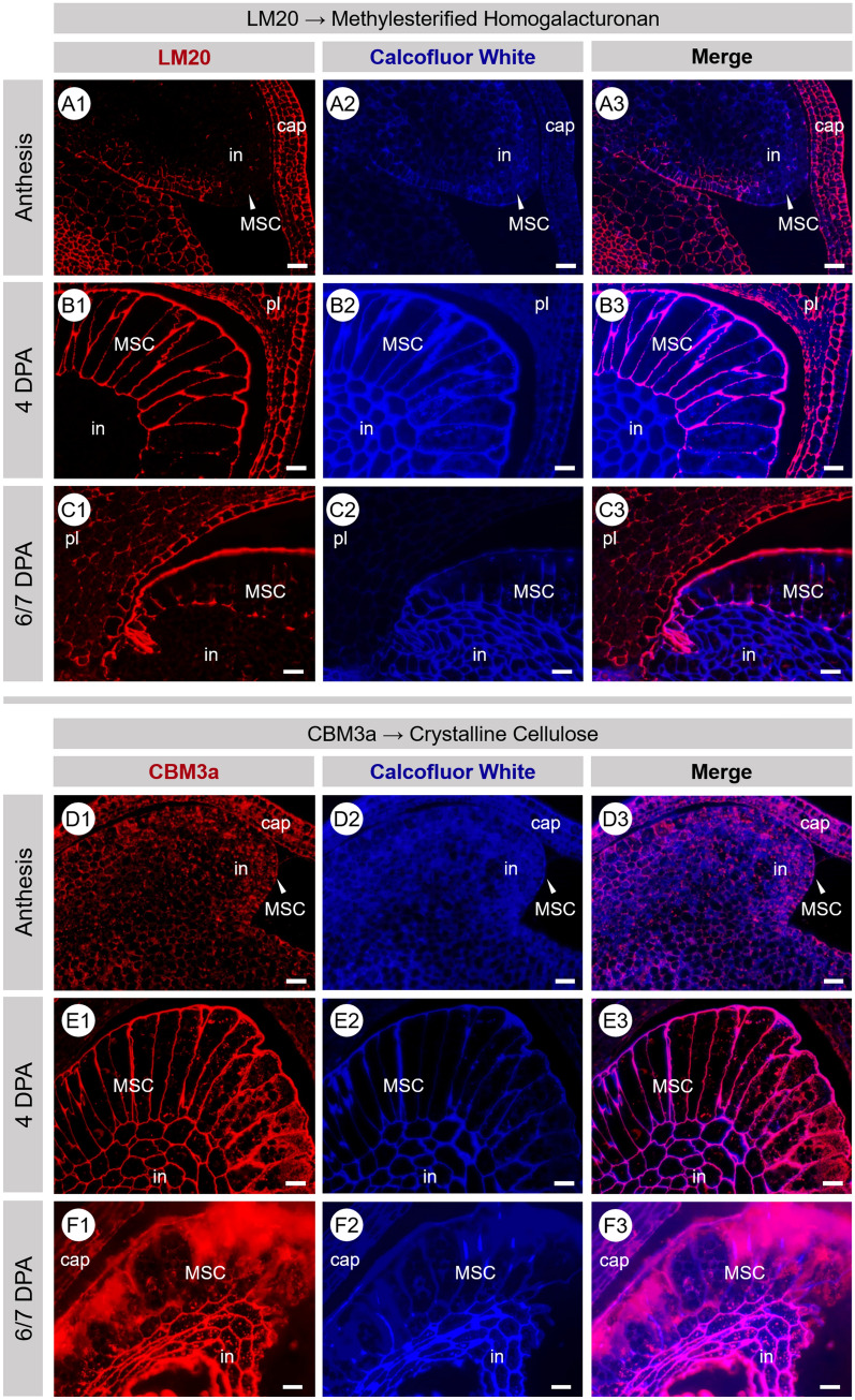
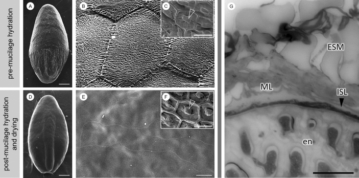
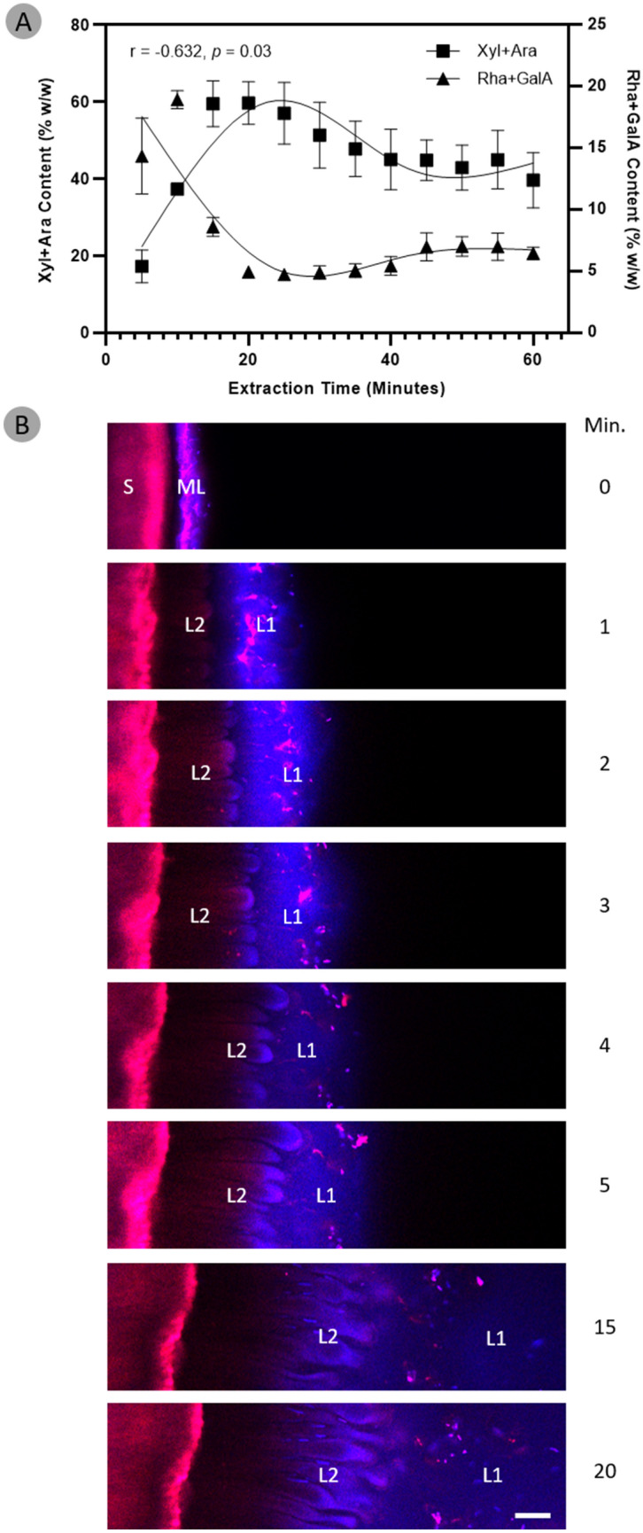
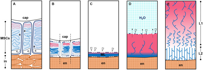
Seed mucilage polysaccharide production, storage and release in Plantago ovata is strikingly different to that of the model plant Arabidopsis. We have used microscopy techniques to track the development of mucilage secretory cells and demonstrate that mature P. ovata seeds do not have an outer intact cell layer within which the polysaccharides surround internal columellae. Instead, dehydrated mucilage is spread in a thin homogenous layer over the entire seed surface and upon wetting expands directly outwards, away from the seed. Observing mucilage expansion in real time combined with compositional analysis allowed mucilage layer definition and the roles they play in mucilage release and architecture upon hydration to be explored. The first emergent layer of hydrated mucilage is rich in pectin, extremely hydrophilic, and forms an expansion front that functions to 'jumpstart' hydration and swelling of the second layer. This next layer, comprising the bulk of the expanded seed mucilage, is predominantly composed of heteroxylan and appears to provide much of the structural integrity. Our results indicate that the synthesis, deposition, desiccation, and final storage position of mucilage polysaccharides must be carefully orchestrated, although many of these processes are not yet fully defined and vary widely between myxospermous plant species.


Figures

Figure 1
Figure 1
(A) The fruit development of P. ovata. Each fruit contains two ovules separated by placental tissue. Plantago species have a circumscissile capsule, also known as a pyxis. The arrow indicates the equator, where the zone of dehiscence is visible, the square bracket highlights the operculum, which detaches during dehiscence, and *indicates the end that joins the fruit to the rachis. Bar 1 mm. (B) A dissected fruit at 13 DPA, showing two immature seeds and in (C) one of the seeds has been further dissected to show the mucilage polysaccharide layer, which has been peeled off the seed and is the remnant of the integument tissue. Longitudinal (D) and transverse (E) cross sections of a developing fruit at 7 DPA, stained with toluidine blue. Em embryo sac, MSC mucilage secretory cells, in integument.
Figure 2
Figure 2
Toluidine blue-stained transverse sections of the developing integument of P. ovata. The sections show the tissues and components that are the: endosperm (en); integument (in); mucilage polysaccharides (μ); mucilage secretory cells (MSCs); mucilage polysaccharide layer (ML); intensely stained layer (ISL); and starch granules (s) at days post-anthesis (DPA). Scale bar 50 μm.
Figure 3
Figure 3
Fluorescence micrographs of transverse sections of developing P. ovata seeds labelled with LM20 and CBM3a (red/pink) at anthesis (A,D), at 4 DPA (B,E) and at 6/7 DPA (C,F). MSC cell walls show strong labelling of highly methylesterified HG (LM20) and crystalline cellulose (CBM3a) that diminishes in intensity and organisation as development/mucilage polysaccharide accumulation continues and/or as MSC cell walls disintegrate. Samples are counter-stained with calcofluor white (blue). Scale 20 μm. DPA days post-anthesis, HG homogalacturonan, MSC mucilage secretory cell, in integument, pl placenta, cap capsule.
Figure 4
Figure 4
Scanning electron micrographs show that P. ovata does not contain a columella (A,B). Inset (C) shows a scanning electron micrograph of the seed surface of Arabidopsis with the columella structure indicated with an arrowhead. In P. ovata, the wrinkled texture of the dry mucilage polysaccharide layer (ML) and hexagonal shapes of the distal MSC wall remnants disappear after mucilage is hydrated and allowed to dry back onto the seed surface, unfixed (D,E), leaving it extremely smooth. This contrasts with Arabidopsis where the distinct columella structure persists and remains clearly visible after the same process (F). Toluidine blue-stained cross sections of the mature seeds fixed in aqueous fixative (G) reveal that the seed mucilage (ESM) expands from the ML, which sits on top of an intensely stained layer (ISL) that separates the mucilage polysaccharide layer from the endosperm. En endosperm. Scales A,D,G 500 μm; B,E,H 50 μm; C,F 30 μm.
Figure 5
Figure 5
(A) Seeds were selected from developmentally-impaired gamma-irradiated P. ovata mutant 69-1 generated previously by Tucker et al.. When these seeds were imbibed in a ruthenium red solution (0.01% w/v) for 10 min at room temperature (B) mucilage expanded from all seeds with different architectures but two typical mucilage layers (L1 and L2) were present. em embryo, en endosperm, ML mucilage layer, WT wild-type. Scale bar 1 mm. (C) Analysis of mucilage yield and composition revealed no significant difference (ns) from the wild-type (p > 0.05, Student’s t test).
Figure 6
Figure 6
The composition and structure of seed mucilage changes over the course of its expansion. (A) Compositional analysis reveals pectin-associated monosaccharides (rhamnose and galacturonic acid) are most abundant during the initial expansion of mucilage and rapidly decrease in concentration thereafter. Heteroxylan-associated monosaccharides (xylose and arabinose) are also present in the initial expansion of mucilage, in almost similar amounts to pectin. Contrasting to pectin, the heteroxylan-associated monosaccharides rapidly increase in concentration and go on to make up the bulk of total expanded mucilage. Data have been fitted with cubic spline curves to highlight trends. (B) The dynamics of seed mucilage expansion in mature P. ovata seeds were observed in real-time over a period of 20 min using confocal microscopy. Seeds were pre-stained with 0.4% Direct Red 23 and 0.1% Calcofluor White. Upon imbibition in water, a sudden “explosion” of an extremely hydrophilic and non-structured mucilage layer emerges (L1). Following L1, a more structured and anemone-like layer of mucilage expands outwards (L2) and by 20 min, L2 has reached its maximal expansion distance and L1 has mostly dissipated into the surrounding aqueous environment. Scale bar 100 μm. S mature seed, ML mucilage polysaccharide layer on dry seed, L1 layer 1, L2 layer 2 N.B. time 0 min is a dry seed that has been pre-stained, water was added after this image was taken.
Figure 7
Figure 7
A proposed model of the polysaccharide deposition and mucilage expansion mechanism in P. ovata. (A) Mucilage polysaccharides pectin (pink) and heteroxylan (blue) are polarly synthesised and deposited into the outer apoplast (a) of mucilage secretory cells (MSCs), (B) which become compressed between the endosperm (en) and capsule wall (cap), obliterating the radial walls and releasing their contents into a (C) continuous laminated cell-free mucilage polysaccharide layer (ML) on the external seed surface when released from the capsule. (D) Upon exposure to an aqueous environment, the hydrophilic mucilage starts to expand outwards from the seed surface. The first layer to expand is rich in extremely hydrophilic and soluble pectin and is topped by fragile remnants of the distal MSC walls (arrowheads) that dissolve as hydration continues. This layer works to provide a hydration cascade to initiate and jumpstart hydration and swelling of the more gel-like, less hydrophilic polymers. (E) A hypothesised distribution of mucilaginous polysaccharides. The pink gradient is indicative of the distribution of pectin which is restricted to the periphery (or ‘mucilage expansion front’) of the expanded seed mucilage (as per Fig. 6) whilst the xylan polysaccharides (blue strands) are evenly distributed (Fischer et al.; Guo et al.; Yu et al.). In L1, the enrichment of pectin may proportionally modulate the solubility/extractability of xylan while in L2, pectin is not present and thus xylan polymers form a more robust, gel-like layer. In integument, c cytoplasm.

References

    1. Phan JL, Burton RA. New insights into the composition and structure of seed mucilage. Annu. Plant Rev. Online. 2018;1:1–41.
    1. Macquet A, Ralet M-C, Kronenberger J, Marion-Poll A, North HM. Insitu, chemical and macromolecular study of the composition of Arabidopsisthaliana seed coat mucilage. Plant Cell Physiol. 2007;48:984–999. - PubMed
    1. Harpaz-Saad S, et al. Cellulose synthesis via the FEI2 RLK/SOS5 pathway and CELLULOSE SYNTHASE 5 is required for the structure of seed coat mucilage in Arabidopsis. Plant J. 2011;68:941–953. - PubMed
    1. Yang B, et al. TRM 4 is essential for cellulose deposition in Arabidopsis seed mucilage by maintaining cortical microtubule organization and interacting with CESA 3. New Phytol. 2019;221:881–895. - PMC - PubMed
    1. Yu L, et al. CELLULOSE SYNTHASE-LIKE A2, a glucomannan synthase, is involved in maintaining adherent mucilage structure in Arabidopsis seed. Plant Physiol. 2014;164:1842–1856. - PMC - PubMed

Publication types