. 2020 Jul 16;10(1):11818.
doi: 10.1038/s41598-020-68874-7.

Genetically modified macrophages accomplish targeted gene delivery to the inflamed brain in transgenic Parkin Q311X(A) mice: importance of administration routes

Affiliations

Genetically modified macrophages accomplish targeted gene delivery to the inflamed brain in transgenic Parkin Q311X(A) mice: importance of administration routes

Matthew J Haney et al. Sci Rep. .

Abstract

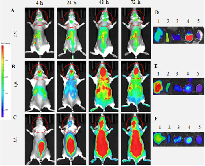
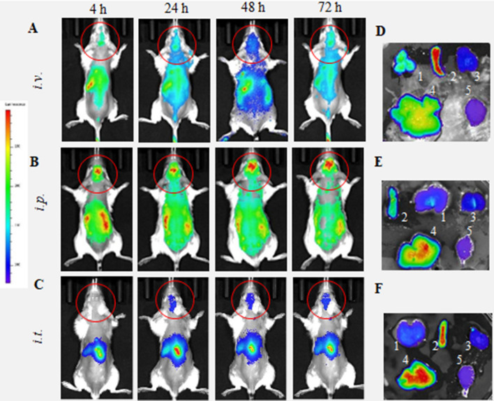
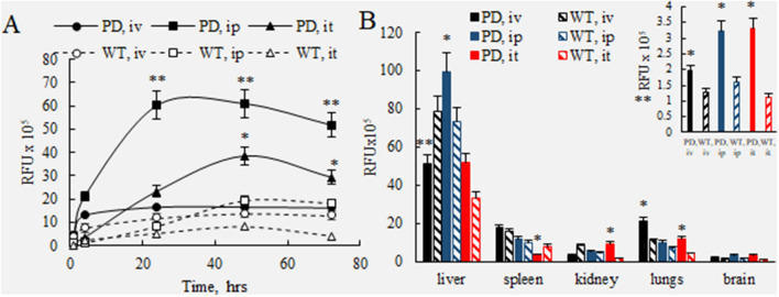
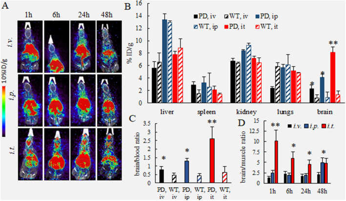
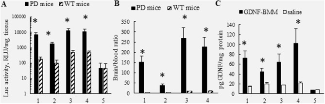
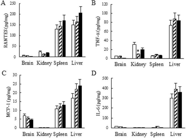
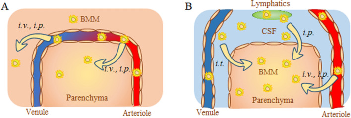
Cell-based drug delivery systems have generated an increasing interest in recent years. We previously demonstrated that systemically administered macrophages deliver therapeutics to CNS, including glial cell line-derived neurotrophic factor (GDNF), and produce potent effects in Parkinson's disease (PD) mouse models. Herein, we report fundamental changes in biodistribution and brain bioavailability of macrophage-based formulations upon different routes of administration: intravenous, intraperitoneal, or intrathecal injections. The brain accumulation of adoptively transferred macrophages was evaluated by various imaging methods in transgenic Parkin Q311(X)A mice and compared with those in healthy wild type littermates. Neuroinflammation manifested in PD mice warranted targeting macrophages to the brain for each route of administration. The maximum amount of cell-carriers in the brain, up to 8.1% ID/g, was recorded followed a single intrathecal injection. GDNF-transfected macrophages administered through intrathecal route provided significant increases of GDNF levels in different brain sub-regions, including midbrain, cerebellum, frontal cortex, and pons. No significant offsite toxicity of the cell-based formulations in mouse brain and peripheral organs was observed. Overall, intrathecal injection appeared to be the optimal administration route for genetically modified macrophages, which accomplished targeted gene delivery, and significant expression of reporter and therapeutic genes in the brain.


Conflict of interest statement

The authors declare no competing interests.

Figures

Figure 1
Figure 1
Biodistribution of DIR-labeled BMM in Parkin Q311(X)A mice by IVIS. DIR-labeled BMM were administered in PD mice (12 mo. of age) (A, D) i.v. (4 × 106 cells/200 µL), (B, E) i.p. (4 × 106 cells/200 µL), or (C, F) i.t. (1 × 106 cells/50 µL). Images were recorded (AC) in live animals at different time points, and (DF) in main organs, liver (1), spleen (2), kidney (3), lungs (4), and brain (5), collected at the endpoint (72 h). Prone representative images show DIR signal accumulation in the brain for all administration routes examined, especially at 24–72 h. Accumulation of labeled macrophages was also observed in the main peripheral organs.
Figure 2
Figure 2
Biodistribution of DIR-labeled BMM in healthy mice by IVIS. DIR-labeled BMM were administered in healthy mice (12 mo. of age) (A, D) i.v. (4 × 106 cells/200 µL), (B, E) i.p. (4 × 106 cells/200 µL), or (C, F) i.t. (1 × 106 cells/50 µL). Images were recorded (AC) in live animals at different time points, and (DF) in main organs, liver (1), spleen (2), kidney (3), lungs (4), and brain (5), collected at the endpoint (72 h). Prone representative images of animals i.v., and i.p. suggest that DIR-BMM accumulate in the brain, although to a much lesser extent than same treatments in Parkin Q311(X)A mice (Fig. 1). Little if any DIR signal was observed in live animals after i.t. administration in healthy animals. Accumulation of labeled macrophages was also observed in the main peripheral organs.
Figure 3
Figure 3
Quantification of BMM accumulation in the main organs of PD and WT mice. Parkin Q311(X)A mice (12 mo. of age) were injected with DIR-BMM using different routes of administration and imaged by IVIS. Wild type mice were used as healthy controls. (A) Quantification of fluorescence levels in the brain area of PD mice (filled symbols) or WT animals (empty symbols) injected with DIR-BMM through i.v. (circles), i.p. (squares), or i.t. (triangles). The highest levels were detected in PD mice with i.p. and i.t. administrations at 24–72 h time frame. Accumulation levels of BMM in PD mouse brain were significantly greater than those in WT littermates. (B) At the endpoint (72 h), mice were sacrificed, perfused, and fluorescent levels of main organs (i.e. liver, spleen, kidney, lungs, and brain) were assessed by IVIS Aura software. The highest BMM accumulation was recorded in liver, spleen, and lungs. The amount of cell-carriers was significantly greater in the brain area of PD mice compared with WT littermates (insert). Values are the means ± SEM (N = 4), *p < 0.05; **p < 0.005 compared with WT healthy mice; #p < 0.05 compared to PD mice injected through i.v. route. Individual data points shown in Supplementary Tables S1 and S2.
Figure 4
Figure 4
Biodistribution kinetics of 64Cu-labeled macrophages in Parkin Q311(X)A and WT mice by PET. Radioactively-labeled BMM were injected into PD or WT mice (12 mo. of age) though i.v., i.p., or i.t. routes (2.5 × 106 cells/50 µL). The animals were imaged by PET over 48 h-time period after the injection (A). At the endpoint (48 h), mice were sacrificed, perfused, and the radioactivity in the blood and main organs was measured. Panels present (A) representative PET images, (B) percentage of the injected dose (%ID/g), (C) brain/blood ratio, and (D) brain/muscle ratio. The greatest brain accumulation of 64Cu-BMM was detected in PD mice with i.t. injections. The amount of 64Cu-BMM accumulated in the PD mouse brain was significantly greater than those in WT animals. Values are the means ± SEM (N = 4), *p < 0.05; **p < 0.005 compared with WT healthy mice (B, C), or i.v. injections (D). Individual data points shown in Supplementary Tables S4–S6.
Figure 5
Figure 5
Biodistribution of 19F-labeled macrophages in Parkin Q311(X)A mice over time by MRI. 19F- labeled BMM were injected i.t. into PD mice (12 mo. of age, 1 × 106 cells/50 µL) and 1H/19F fusion image was obtained. Anatomic image is represented in gray scale with cell loading represented by the color. A phantom standard is external to the animal. Representative dual mode brain MR images show significant accumulation of i.t. administered BMM in the brain, especially at 48 h time point.
Figure 6
Figure 6
Reporter and therapeutic gene expression in brain of Parkin Q311(X)A mice after single i.t. injection of pre-transfected BMM. (A) Luc-transfected BMM (A, B) were injected through i.t. route (2.5 × 106 cells/50 µL) to PD mice (black bars) or WT mice (striped bars). Mice were sacrifices 48 h after injection, perfused, brains were harvested and luciferase activity was measured in different brain regions [i.e. frontal cortex (1), midbrain (2), cerebellum (3), and pons (4)), and blood (5)]. Luciferase activity in each brain region was significantly greater in PD mouse brain regions than those in WT counterparts (A). Total brain/blood ratio of luciferase activity in PD mice was 25 times greater than in WT mice (B), indicating that BMM target inflamed brain tissues. (C) PD mice were injected with the same amount of GDNF-transfected BMM through i.t. route (black bars), or saline (white bars). Mice were sacrifices 48 h after injection, perfused, brains were harvested and GDNF expression was measured in different brain regions by ELISA. GDNF levels in each brain region were greater in PD mouse brain regions injected with GDNF-BMM than controls injected with saline. Negligible differences in GDNF levels in the blood were observed. Values are means ± SEM (N = 6). *p < 0.05 compared to WT healthy mice (A, B), or saline injected control mice (C). Individual data points shown in Supplementary Tables S8–S10.
Figure 7
Figure 7
Effect of BMM administration on the levels of inflammatory signals in mice. Healthy WT mice were injected with saline (white bars), sham BMM (stripped bars), or GDNF-transfected BMM (black bars), and the inflammation signals in the main organs was assessed by the levels of RANTES (A), TNF-a (B), MCP-1 (C), IL-6 (D). No significant increases of inflammatory signals were found in the brain. Some slight increases in RANTES, and MCP-1 were registered in spleen and liver (A, C). TNF-a levels were significantly decreased in spleen upon sham BMM injection. Values are means ± SEM (N = 10). *p < 0.05 compared to saline-injected control mice. Individual data points shown in Supplementary Tables S11–S14.
Figure 8
Figure 8
Schematic representation of different pathways for adoptively transferred macrophages to the brain. Autologous macrophages transfected with reporter or therapeutic—encoding pDNA were injected via i.v., i.p., or i.t. routes. Different ways of brain entrance across (A) the BBB, or (B): CSF barrier are shown. (A) Macrophages injected through i.v. or i.p. routes cross the BBB and deliver the encoded proteins to the parenchyma. (B) In addition, macrophages injected i.p. may enter the brain through lymphatic system. Macrophages administered through i.t. route mainly enter through the CSF barrier.

References

    1. Malek N, Swallow DMA, Grosset KA, Lawton MA, Marrinan SL, Lehn AC, Bresner C, Baiai N, Baker RA, Ben-Shlomo Y, Burn DJ, Foltynie T, Hardy J, Morris HR, Williams NM, Wood N, Grosset DG. Tracking Parkinson’s: study design and baseline patient data. J Parkinsons Dis. 2015;5:947–959. - PMC - PubMed
    1. Farrer MJ. Genetics of Parkinson disease: paradigm shifts and future prospects. Nat. Rev. Genet. 2006;7:306–318. - PubMed
    1. Gurme ST, Surwase SN, Patil SA, Jadhav SB, Jadhav JP. Optimization of biotransformation of l-tyrosine to l-DOPA by Yarrowia lipolytica-NCIM 3472 using response surface methodology. Indian J. Microbiol. 2013;53:194–198. - PMC - PubMed
    1. Wang Q, Liu Y, Zhou J. Neuroinflammation in Parkinson’s disease and its potential as therapeutic target. Transl. Neurodegener. 2015;4:19. - PMC - PubMed
    1. McGeer PL, Itagaki S, Boyes BE, McGeer EG. Reactive microglia are positive for HLA-DR in the substantia nigra of Parkinson’s and Alzheimer’s disease brains. Neurology. 1988;38:1285–1291. - PubMed

Publication types