. 2021 Mar;14(2):491-499.
doi: 10.1038/s41385-020-0322-6. Epub 2020 Jul 16.

Mycobacterium tuberculosis-specific CD4 T cells expressing CD153 inversely associate with bacterial load and disease severity in human tuberculosis

Affiliations

Mycobacterium tuberculosis-specific CD4 T cells expressing CD153 inversely associate with bacterial load and disease severity in human tuberculosis

Elsa Du Bruyn et al. Mucosal Immunol. 2021 Mar.

Abstract

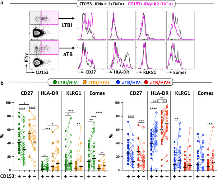
Recent data from mice and non-human primate models of tuberculosis suggested that CD153, a TNF super family member, plays an important role in Mycobacterium tuberculosis (Mtb) control. However, this molecule has not been comprehensively evaluated in humans. Here, we show that the proportion of Mtb-specific CD4 T cells expressing CD153 was significantly reduced in active TB patients compared to latently infected persons. Importantly, the CD153+ Mtb-specific CD4 response inversely correlated with lung bacterial load, inferred by Xpert cycle threshold, irrespective of HIV status. Antitubercular treatment partially restored CD153 expression on Mtb-specific CD4 T cells. This is the first report of a subset of Mtb-specific CD4 T cells showing strong negative correlation with bacterial burden. Building on substantial evidence from animal models implicating CD153 as a mediator of host protection, our findings suggest it may play a similar role in humans and its measurement may be useful to evaluate TB vaccine efficacy.


Conflict of interest statement

The authors declare no competing interests.

Figures

Fig. 1
Fig. 1. Sputum Xpert MTB/RIF cycle threshold (Xpert CT) values associates with other measures of Mtb Burden (time to Mtb culture positivity (TTP) and smear microscopy grade), systemic inflammation (CRP), and radiographic evidence of disease severity (Timika score).
a Correlation between Xpert CT values and time to Mtb culture positivity in HIV-uninfected (blue) and HIV-infected (red) participants. b Correlation between smear microscopy grade and Xpert CT values. Horizontal lines represent the median values. c, d Correlations between Xpert CT values and plasma C-reactive protein and Timika score on chest radiograph. The solid lines indicate the linear regression and correlations between measures were performed using a two-tailed nonparametric Spearman test.
Fig. 2
Fig. 2. The proportion of Mtb300-specific CD4 T cells expressing CD153 is reduced in aTB compared to LTBI regardless of HIV status.
a Representative flow cytometry plots showing the expression of CD153 in Mtb300-spcific CD4 T cells in one LTBI and one aTB participant. No stim = no stimulation; Mtb300 = stimulation with a pool of 300 Mtb-derived peptides. b Proportion of Mtb300-specific CD4 T cells expressing CD153 in LTBI/HIV− (n = 35, green), aTB/HIV− (n = 33, blue), LTBI/HIV+ (n = 30, orange), and aTB/HIV+ (n = 35, red). Error bars indicate medians and interquartile ranges. Statistical comparisons were performed using One-way ANOVA Kruskal–Wallis test with a Dunn’s multiple comparison test. CD153 expression was assessed only on Mtb300-specific response (i.e CD4 T cells expressing any measured cytokines) exhibiting more than 30 events. c CD153 expression in Mtb300-specific CD4 T cells in LTBI and aTB HIV-infected participants stratified based on plasma HIV viral load (aviremic vs viremic). Median VL for each subgroup is presented at the bottom of the graph. Statistical comparisons were performed using Mann–Whitney test. d Correlation between HIV viral load and the proportion of CD153+ Mtb300-specific CD4 T cells in the aTB (top) and LTBI (bottom) groups. Correlations were tested by a two-tailed nonparametric Spearman rank test.
Fig. 3
Fig. 3. Polyfunctional Mtb300-specific CD4 T cells (i.e CD153+ IL-2+ IFNγ+ TNFα+) are selectively reduced in aTB participants compared to LTBI, regardless of HIV status.
The x-axis displays each of the different response patterns, the composition of which is denoted with a dot for the presence of CD153, IL‐2, IFNγ, and TNFα. The proportion of each response pattern contributing to the total Mtb300-specific CD4 response per individual is shown. The median (gray bar) and interquartile ranges (box) are shown. Subsets accounting for <1% of the total Mtb300-specific CD4 T cell response are not displayed. Each response pattern is color‐coded, and data are summarized in the pie charts, where each pie slice represents the median contribution of each response pattern to the total Mtb300 response. A Wilcoxon rank-sum test was used to compare response patterns between groups (****P < 0.0001, ***P < 0.001, **P < 0.01). Statistical differences between pie charts were defined using a permutation test.
Fig. 4
Fig. 4. The proportion of CD153+ Mtb300-specific CD4 T cells inversely correlates with Mtb bacterial burden, irrespective of HIV status.
a Correlation between Xpert CT values and the proportion of CD153+ Mtb300-specific CD4 T cells in HIV-uninfected (blue) and HIV-infected (red) persons. Linear regression and 95% confidence band are depicted. b Correlation between Xpert CT values and the proportion of IL-2+ Mtb300-specific CD4 T cells in HIV-uninfected and HIV-infected persons. c Correlation between the proportion of CD153+ Mtb300-specific CD4 T cells and the proportion of IL-2+ Mtb300-specific CD4 T cells in HIV-uninfected and HIV-infected persons. Correlations were tested by a two-tailed nonparametric Spearman rank test.
Fig. 5
Fig. 5. CD153+ polyfunctional Mtb300-specific CD4 T cells are phenotypically less activated and differentiated than CD153− polyfunctional cells.
a Representative flow cytometry overlay plots of CD27, HLA-DR, KLRG1, and Eomes expression in CD153+ IL-2+ IFNγ + TNFα+ (pink) and CD153− IL-2+ IFNγ+ TNFα+ (black) Mtb300-specific CD4 T cells in one LTBI and one aTB participant. b Summary graph of the expression of CD27, HLA-DR, KLRG1, and Eomes in CD153+ IL-2+ IFNγ+ TNFα+ [CD153+] and CD153- IL-2+ IFNγ+ TNFα+ [CD153−] Mtb300-specific CD4 T cells in LTBI (left) and aTB (right) participants. Only CD153+ IL-2+ IFNγ+ TNFα+ and CD153- IL-2+ IFNγ+ TNFα+ subpopulations with more than 30 events were considered for phenotyping. Statistical comparisons were performed using a paired nonparametric Wilcoxon test. (****P < 0.0001, ***P < 0.001, **P < 0.01, *P < 0.05).
Fig. 6
Fig. 6. Partial restoration of CD153+ Mtb300-specific CD4 T cells after antitubercular therapy (ATT).
a Comparison of the proportion of CD153-expressing Mtb300-specific CD4 T cells before (baseline, BL) and after 24 weeks of TB treatment (W24) in HIV- uninfected and HIV-infected patients. Statistical comparisons were performed using a paired nonparametric Wilcoxon test for longitudinal data and a Mann–Whitney test between groups. b Fold change in CD153+ Mtb300-specific CD4 T cells between BL and W24. Statistical comparison was performed using a nonparametric Mann–Whitney test. c Polyfunctional profile of Mtb300 CD4 responses pre and post ATT. The x-axis displays each response pattern, the composition of which is denoted with a dot indicative of the presence of CD153, IL-2, IFNγ, and TNFα. The median (gray bar) and interquartile ranges (box) are shown. Subsets accounting for less than 1% of the total Mtb300-specific CD4 T cell response are not displayed. Each response pattern is color‐coded, and data are summarized in the pie charts. A Wilcoxon rank-sum test was used to compare response pattern between groups (****P < 0.0001, ***P < 0.001, **P < 0.01). Statistical differences between pie charts were defined using a permutation test.

References

    1. WHO (World Health Organization). Global Tuberculosis Report. (WHO, 2019). https://www.who.int/tb/publications/global_report/en/.
    1. Flynn JL, et al. An essential role for interferon gamma in resistance to Mycobacterium tuberculosis infection. J. Exp. Med. 1993;178:2249–2254. - PMC - PubMed
    1. Moreira-Teixeira L, Mayer-Barber K, Sher A, O’Garra A. Type I interferons in tuberculosis: foe and occasionally friend. J. Exp. Med. 2018;215:1273–1285. - PMC - PubMed
    1. Lalvani A, Millington KA. T cells and tuberculosis: beyond interferon-gamma. J. Infect. Dis. 2008;197:941–943. - PubMed
    1. Esmail H, et al. The immune response to Mycobacterium tuberculosis in HIV-1-coinfected persons. Annu Rev. Immunol. 2018;36:603–638. - PubMed

Publication types