. 2020 Jul 16;10(1):11815.
doi: 10.1038/s41598-020-68867-6.

Validation of virtual reality orbitometry bridges digital and physical worlds

Affiliations

Validation of virtual reality orbitometry bridges digital and physical worlds

Peter M Maloca et al. Sci Rep. .

Abstract

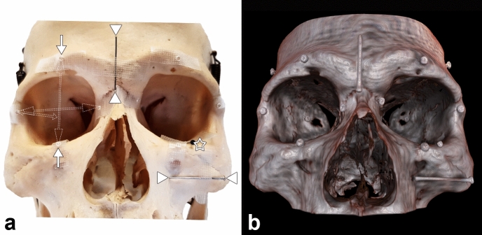
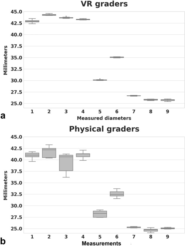
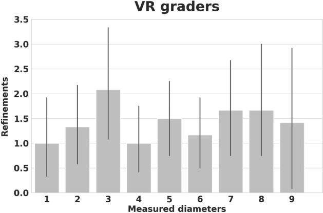
Clinical science and medical imaging technology are traditionally displayed in two dimensions (2D) on a computer monitor. In contrast, three-dimensional (3D) virtual reality (VR) expands the realm of 2D image visualization, enabling an immersive VR experience with unhindered spatial interaction by the user. Thus far, analysis of data extracted from VR applications was mainly qualitative. In this study, we enhance VR and provide evidence for quantitative VR research by validating digital VR display of computed tomography (CT) data of the orbit. Volumetric CT data were transferred and rendered into a VR environment. Subsequently, seven graders performed repeated and blinded diameter measurements. The intergrader variability of the measurements in VR was much lower compared to measurements in the physical world and measurements were reasonably consistent with their corresponding elements in the real context. The overall VR measurements were 5.49% higher. As such, this study attests the ability of VR to provide similar quantitative data alongside the added benefit of VR interfaces. VR entails a lot of potential for the future research in ophthalmology and beyond in any scientific field that uses three-dimensional data.


Conflict of interest statement

The research of PCC, BF, MZ, and CJ was supported by the Werner Siemens Foundation through the MIRACLE project. PCC is the owner of the described method. All other authors have no competing interests.

Figures

Figure 1
Figure 1
Diameter measurements of physical world compared to digital VR world of the human orbit. Two different groups of measuring landmarks were placed on a physical skull. (a) For each orbit, six small metal beads were placed in the three main axes, defining three different diameters (for better overview, these were illustrated only on the right orbit; single arrows representing the longitudinal beads, double headed arrows indicate the measured longitudinal and frontal diameter measured from the outside border; short double arrow indicates the oblique and lateral diameter inside the orbit). In addition, three metal pins (arrow head) were attached in the aforementioned axes on the skull near the orbit, whereby only the head of the third nail (asterisk) is visible from outside. VR imaging (b) of the identical skull from (a) displays the corresponding landmarks. The osseous structures are rendered realistically in VR, whereas the metal particles appeared somewhat enlarged. The metallic objects are relatively sharp-edged in VR. No image degradation has been noted, indicating that the used landmarks and the scan protocol parameters during CT acquisition were appropriate. Abbreviations: computed tomography (CT); virtual reality (VR). The figure was created using Adobe Photoshop 2020 (Adobe Inc., San Jose, US, licence 65230035) and image b was obtained from the described software SpectoVR (version 3.1.0, Diffuse ltd., Heimberg, Switzerland, https://www.diffuse.ch).
Figure 2
Figure 2
Results of physical diameter measurements compared to digital VR world measurements. In both worlds, it was possible to measure all diameters. In the physical world (a), using a caliper, the deviation appeared higher when compared to VR. The oblique diameter (location # 3) was not optimally measurable. In contrast, the pins that were placed in relatively easily accessible and visually controllable locations showed significantly less variation. In the VR (b), the results are consistent, irrespective of the location.
Figure 3
Figure 3
This Bland–Altman plot visualizes the agreement between the physical and virtual measuring method. An average difference of 2 mm (solid line) is observed. The differences lie within the 95% confidence interval (dashed lines) with one exception.
Figure 4
Figure 4
Refinement bar plots of diameter measurements in virtual reality (VR). Because the VR users were always digitally tracked, it was possible to record and display their performance instantly: An additional parameter is offered by VR by determining how often a measurement had been corrected before it had been saved. The mean overall correction of all graders was 1.5. This is not quantifiable in the physical world and may serve as a novel benchmark regarding the performance of a VR user.

References

    1. Rootman J. Diseases of the Orbit: A Multidisciplinary Approach. Philadelphia: Lippincott Williams & Wilkins; 2003.
    1. Shields JA, Shields CL, Scartozzi R. Survey of 1264 patients with orbital tumors and simulating lesions: The 2002 montgomery lecture, part 1. Ophthalmology. 2004;111:997–1008. doi: 10.1016/j.ophtha.2003.01.002. - DOI - PubMed
    1. Shinder R, Al-Zubidi N, Esmaeli B. Survey of orbital tumors at a comprehensive cancer center in the United States. Head Neck. 2011;33:610–614. doi: 10.1002/hed.21498. - DOI - PubMed
    1. Smolarz-Wojnowska A, Essig H, Gellrich NC, Klein CM. Orbital tumours in children and adolescents. Differential diagnostics and clinical symptoms. Der Ophthalmol. Zeitschrift der Deutschen Ophthalmol. Gesellschaft. 2010;107:543–548. doi: 10.1007/s00347-009-2068-0. - DOI - PubMed
    1. Marino M, et al. Orbital diseases mimicking graves' orbitopathy: A long-standing challenge in differential diagnosis. J. Endocrinol. Investig. 2020;43:401–411. doi: 10.1007/s40618-019-01141-3. - DOI - PubMed

Publication types