. 2020 Aug 25;14(8):9347-9363.
doi: 10.1021/acsnano.0c03981. Epub 2020 Jul 22.

Nanofiber Technology for Regenerative Engineering

Affiliations

Nanofiber Technology for Regenerative Engineering

Kenneth S Ogueri et al. ACS Nano. .

Abstract

Regenerative engineering is powerfully emerging as a successful strategy for the regeneration of complex tissues and biological organs using a convergent approach that integrates several fields of expertise. This innovative and disruptive approach has spurred the demands for more choice of biomaterials with distinctive biological recognition properties. An ideal biomaterial is one that closely mimics the hierarchical architecture and features of the extracellular matrices (ECM) of native tissues. Nanofabrication technology presents an excellent springboard for the development of nanofiber scaffolds that can have positive interactions in the immediate cellular environment and stimulate specific regenerative cascades at the molecular level to yield healthy tissues. This paper systematically reviews the electrospinning process technology and its utility in matrix-based regenerative engineering, focusing mainly on musculoskeletal tissues. It briefly outlines the electrospinning/three-dimensional printing system duality and concludes with a discussion on the technology outlook and future directions of nanofiber matrices.

Keywords: 3D-printed scaffold; biodegradable polymer; biomaterial−cell interactions; drug delivery; dual-scale matrix; electrospinning; electrospun nanofiber; stem cells; sustained release; tissue regeneration.


Conflict of interest statement

The authors declare no competing financial interest.

Figures
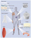
Figure 1.
Figure 1.
The organization of collagen nanofibers found in different musculoskeletal tissues. The tendon and ligament tissues are composed of collagen nanofibers that are uniaxially aligned and wavy. Collagen fibers showed aligned-to-random orientations at tendon-to-bone insertion sites and circumferential alignment in the meniscus and annulus fibrosus. Collagen fibers in the cartilage tissues are arranged in layers. Reprinted with permission from ref 13. Copyright 2013 Future Medicine Ltd.
Figure 2.
Figure 2.
(A) Diagram showing the hierarchical macrostructure constructs of bone along with the cylindrical shape of osteon construct, and microstructure/nanostructure of collagen. Reprinted with permission from ref 14. Copyright 2018 MDPI. (B) Schematics of cell-nanofiber interactions showing the attachment of cell cytoskeleton to the fibers with the aid of integrins. The anisotropic arrangement and alignment of the fibers influence the morphology of the cells and matrix stiffness regulate the cellular migration (durotaxis) as the cells exhibit contraction when in contact with soft region of the matrix. Reprinted with permission from ref 44. Copyright 2017 Elsevier.
Figure 3.
Figure 3.
Schematic diagram of electrospinning process with static and rotating collectors. Randomly-oriented nanofibers are collected on the static collector, while the rotating drum produces uniaxially aligned nanofibers. Reprinted with permission from ref 30. Copyright 2018 Georg Thieme Verlag KG.
Figure 4.
Figure 4.
Diagrammatic illustrations of coaxial, mixing and multilayering electrospinning systems (A) coaxial electrospinning involving the extrusion of two different polymeric solutions through the inner and outer needle coaxial spinneret (B and C) Images of transmission electron microscopy for coaxially electrospun nanofibers (D) The microscopic view of hollow nanofibers. (E) Mixed and multilayered nanofibers using mixing and multilayering electrospinning process respectively. Reprinted with permission from ref 37. Copyright 2017 Springer Nature.
Figure 5.
Figure 5.
Electrospun nanofibers with aligned-to-random orientations (A) Experimental setup employed in fabricating aligned-to-random nanofiber scaffolds (B) SEM images showing the random and uniaxial aligned PLGA with the random orientation on the left and aligned orientation on the right (C-D) Random and aligned fibers with high magnifications views (E-G) Mechanical properties of the aligned and random nanofiber scaffolds. Aligned nanofibers exhibited higher modulus and ultimate stress. Reprinted with permission from ref 46. Copyright 2009 Royal Society of Chemistry.
Figure 6.
Figure 6.
Effects of fiber diameters on the cellular activities and mechanical properties of scaffolds (A) SEM images of nano and microfibers with different diameters (B) Live/dead cell viability assay on the scaffolds (C) Time-dependent changes of cell aspect ratios. (D) Proliferation of cells with culture time (E) the changes in tensile properties with varying diameters. Reprinted with permission from ref 47. Copyright 2013 Mary Ann Liebert, Inc.
Figure 7.
Figure 7.
Electrospun nanofibers of PLLA (A) SEM image of aligned nanofiber (B) randomly-oriented nanofiber scaffold (C-F) hematoxylin and eosin staining histology of nanofiber-induced tissue formation (G-J) Masson trichrome staining showing the formation of collagen fibers on the aligned and random fibrous scaffolds. Reprinted with permission from ref 51. Copyright 2010 Elsevier.
Figure 8.
Figure 8.
Convergence of 3D printing and electrospinning Technologies (A) Fabrication of dual-scale scaffold with meshes (mesh densities of 15s, 30s, 45s, & 120s) evenly distributed throughout the scaffold by electrospinning onto the scaffold at appropriate layer during printing (B) SEM images of a 3D-printed scaffold and dual-scale scaffold with electrospun nanofibers. The dual-scale scaffold is obtained when nanofibers are electrospun directly onto the scaffold during printing [scale bar = 300μm]. Reprinted with permission from ref 59. Copyright 2020 Mary Ann Liebert, Inc. (C) Photo and SEM images of 3D-printed scaffold and 3D composite scaffold. The composite scaffold is obtained by infusing the dispersed nanofibers into the meshes of 3D-printed scaffold [scale bar = 1mm]. Reprinted with permission from ref 58. Copyright 2016 Royal Society of Chemistry.
Figure 9.
Figure 9.
Schematic diagram showing different modification methods used to integrate bioactive molecules to nanofibers (A) treatment by plasma (B) Immobilization of bioactive molecules by surface graft polymerization (C) co-electrospinning of substrate and bioactive agents. Reprinted with permission from ref 61. Copyright 2009 Elsevier.
Figure 10.
Figure 10.
Preparation of three-dimensional (3D) electrospun nanofibrous scaffold for the regeneration bone tissues using a rat cranial bone defect model. Reprinted with permission from ref 67. Copyright 2019 Elsevier.

References

    1. Laurencin CT; Nair LS Regenerative Engineering: Approaches to Limb Regeneration and Other Grand Challenges. Regener. Eng. Transl. Med 2015, 1 , 1–3. - PMC - PubMed
    1. Laurencin CT; McClinton A Regenerative Cell-Based Therapies: Cutting Edge, Bleeding Edge, and Off the Edge. Regener. Eng. Transl. Med 2020, 6, 1–12. - PMC - PubMed
    1. Ogueri KS; Laurencin CT Matrix-Based Bone Regenerative Engineering In Encyclopedia of Bone Biology, Zaidi M, Ed. Academic Press: Oxford, 2020; pp 135–148.
    1. Ogueri KS; Allcock HR; Laurencin CT Generational Biodegradable and Regenerative Polyphosphazene Polymers and their Blends with Poly (Lactic-Co-Glycolic Acid). Prog. Polym. Sci 2019, 98, 101146. - PMC - PubMed
    1. Ogueri KS; Jafari T; Ivirico JLE; Laurencin CT Polymeric Biomaterials for Scaffold-Based Bone Regenerative Engineering. Regener. Eng. Transl. Med 2018, 5, 128–154. - PMC - PubMed

Publication types

Substances