. 2020 Aug 6;182(3):578-593.e19.
doi: 10.1016/j.cell.2020.06.031. Epub 2020 Jul 16.

Cell Types Promoting Goosebumps Form a Niche to Regulate Hair Follicle Stem Cells

Affiliations

Cell Types Promoting Goosebumps Form a Niche to Regulate Hair Follicle Stem Cells

Yulia Shwartz et al. Cell. .

Abstract

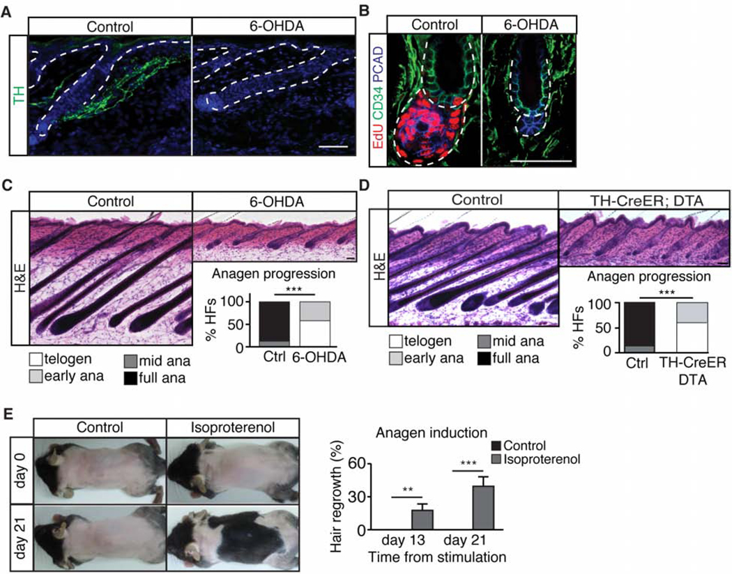
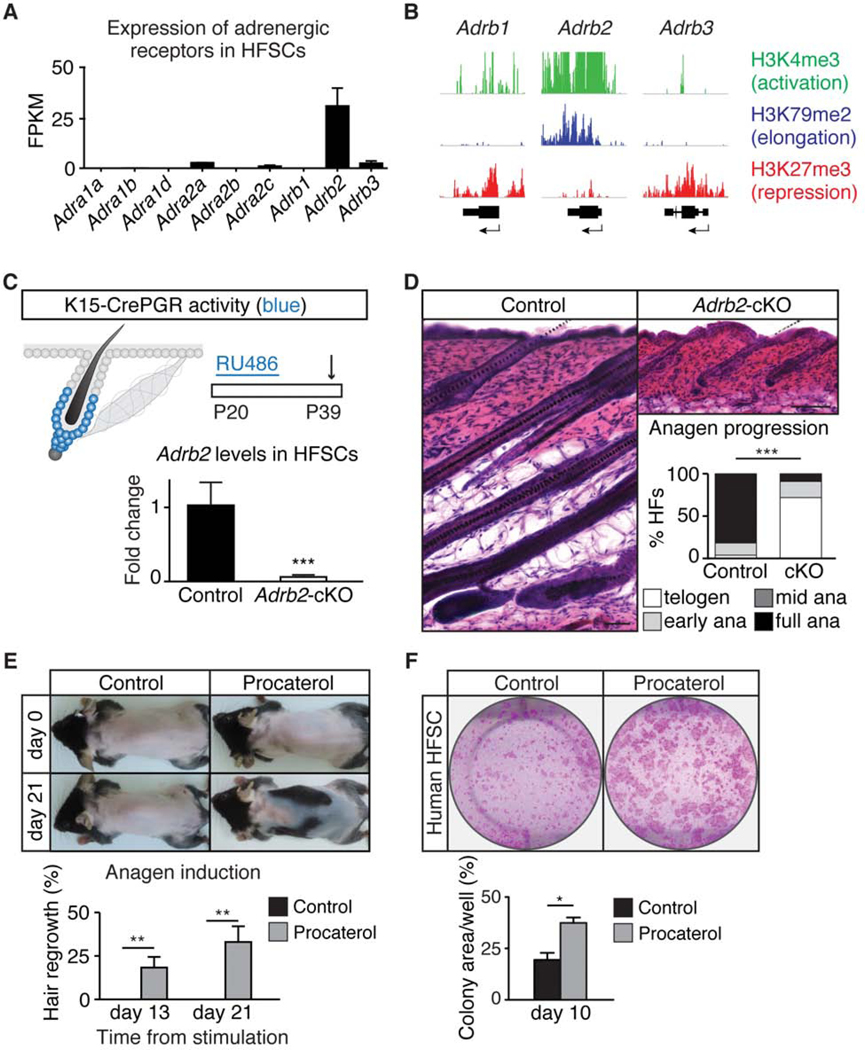
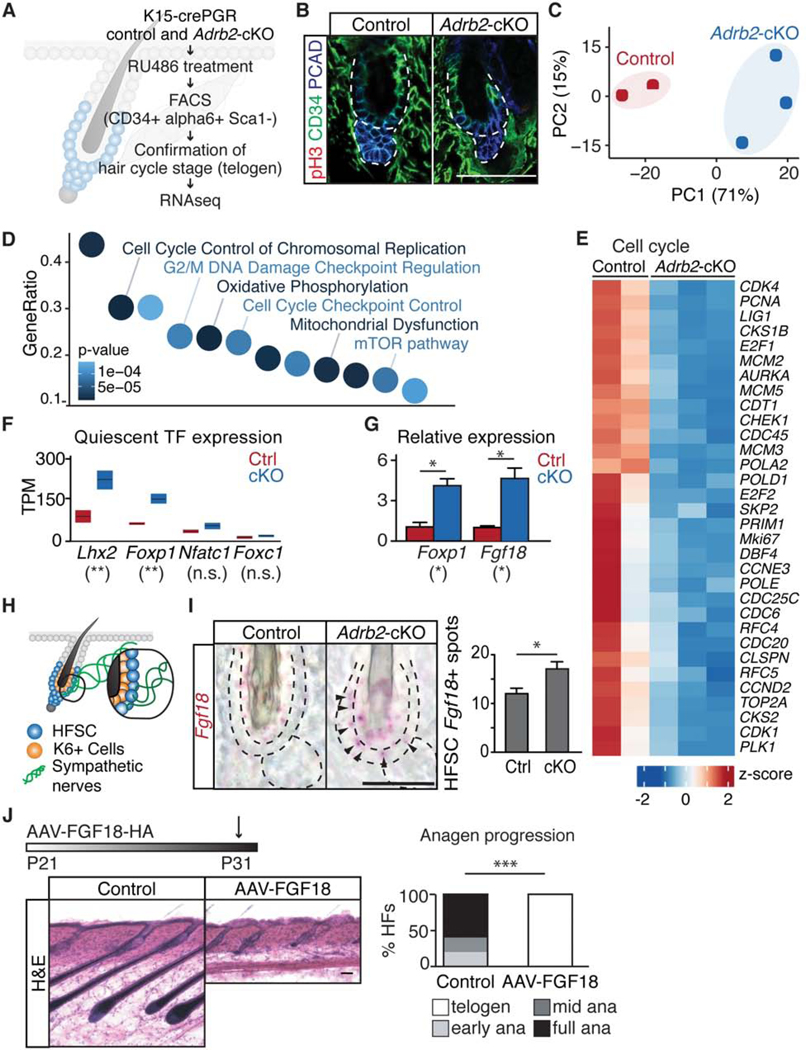
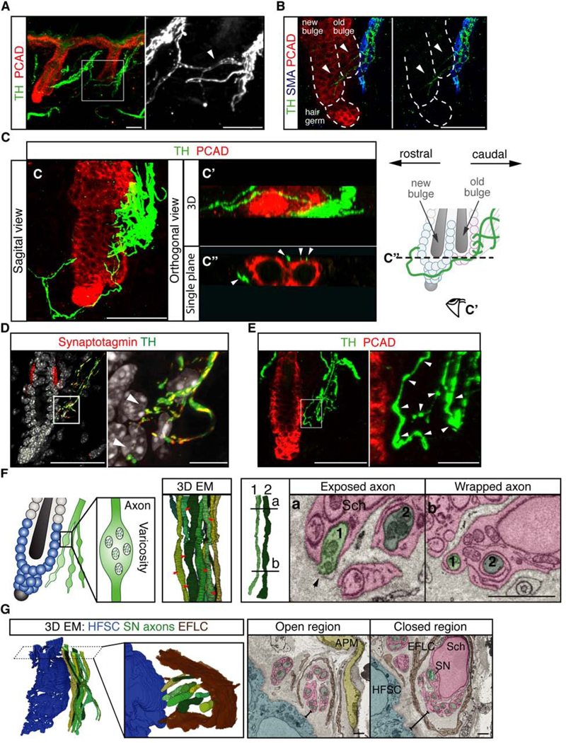
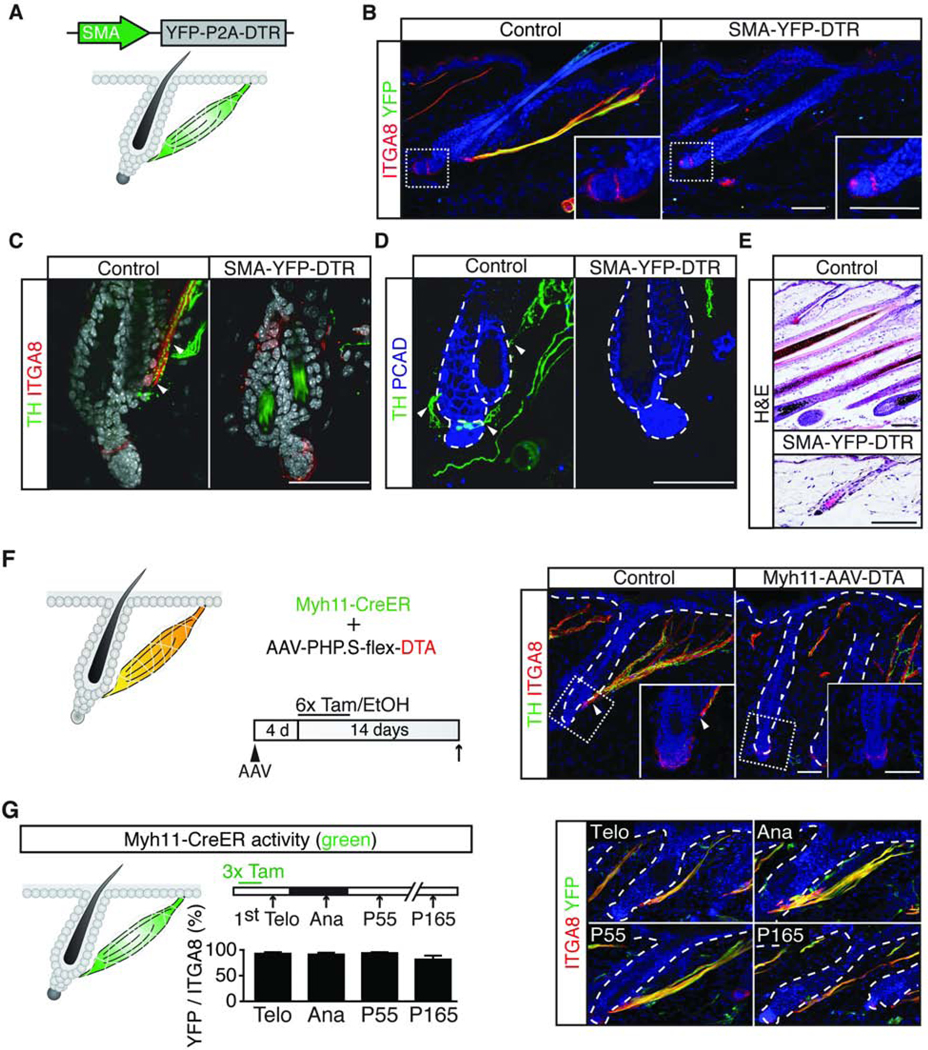
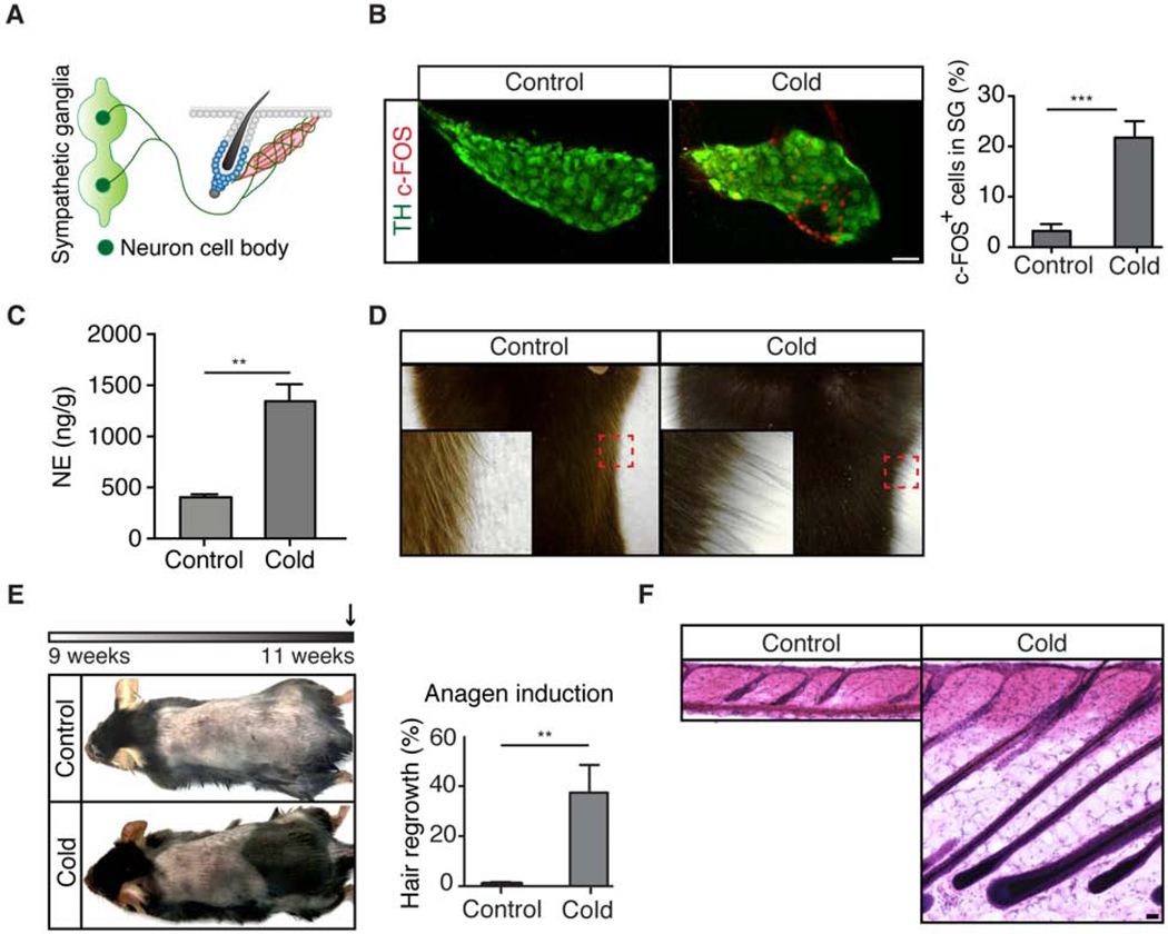
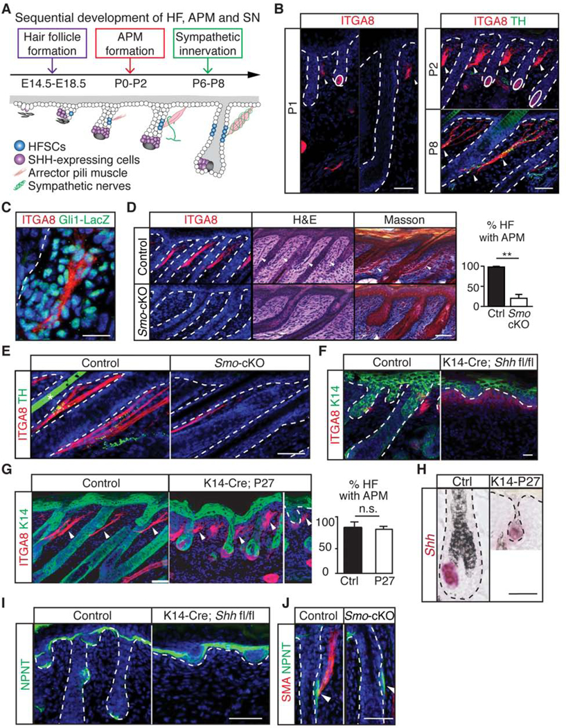
Piloerection (goosebumps) requires concerted actions of the hair follicle, the arrector pili muscle (APM), and the sympathetic nerve, providing a model to study interactions across epithelium, mesenchyme, and nerves. Here, we show that APMs and sympathetic nerves form a dual-component niche to modulate hair follicle stem cell (HFSC) activity. Sympathetic nerves form synapse-like structures with HFSCs and regulate HFSCs through norepinephrine, whereas APMs maintain sympathetic innervation to HFSCs. Without norepinephrine signaling, HFSCs enter deep quiescence by down-regulating the cell cycle and metabolism while up-regulating quiescence regulators Foxp1 and Fgf18. During development, HFSC progeny secretes Sonic Hedgehog (SHH) to direct the formation of this APM-sympathetic nerve niche, which in turn controls hair follicle regeneration in adults. Our results reveal a reciprocal interdependence between a regenerative tissue and its niche at different stages and demonstrate sympathetic nerves can modulate stem cells through synapse-like connections and neurotransmitters to couple tissue production with demands.

Keywords: Adrb2; hair follicle stem cells; nerve-stem-cell interaction; niche; stem cell quiescence; sympathetic nerve.


Conflict of interest statement

Declaration of Interests A provisional patent application has been filed by the President and Fellows of Harvard College based on this work.

Figures

Figure 1.
Figure 1.. Sympathectomy delays anagen entry whereas elevation of sympathetic tone drives anagen entry.
(A) Immunofluorescent staining for tyrosine hydroxylase (TH) in control and sympathectomized (6-OHDA) skin. (B) Immuno-colocalization of EdU, CD34, and P-Cadherin (PCAD) in control and sympathectomized P25 skin. (C) Hematoxylin & Eosin (H&E) staining of control and sympathectomized skin. Graph: hair cycle distribution at P30 (n = 4 – 5 mice per condition, 20 hair follicles (HF) per mouse). (D) H&E staining of control and sympathectomized TH-CreER; Rosa-lsl-attenuated DTA (TH-CreER; DTA) skin. Graph: hair cycle distribution at P31 - P34 (n = 4 – 5 mice per condition, 10 HF per mouse). (E) Topical application of isoproterenol at the 2nd telogen results in precocious anagen entry (n = 10 mice per condition). Graph: back skin hair regrowth (%). Unless otherwise specified, all scale bars = 50 μm. Data are mean ± SEM. *: p<0.05; **: p<0.01; ***: p <0.001. See also Figure S1.
Figure 2.
Figure 2.. HFSC activity is modulated by ADRB2.
(A) Expression of adrenergic receptors in HFSCs (RNA-seq). (B) Chromatin modifications around the loci of Adrb genes in HFSCs. (C) Schematic of K15-CrePGR activity (blue) and experimental design (arrow denotes harvesting). qRT-PCR of Adrb2 from FACS-purified HFSCs of control and K15-CrePGR; Adrb2 fl/fl (Adrb2-cKO) mice (n = 3 mice per condition). (D) H&E staining of control and Adrb2-cKO skin. Graph: hair cycle distribution (n = 5 – 7 mice per condition, 20 HF per mouse). (E) Topical application of procaterol (ADRB2 agonist) drives premature anagen entry (n = 10 mice per condition). Graph: back skin hair regrowth (%). (F) Colony formation assay on control and procaterol-treated human HFSCs. Graph: area covered by colonies (n = 3 – 5 wells per condition). Data are mean ± SEM. *: p<0.05; **: p<0.01; ***: p <0.001. See also Figure S2.
Figure 3.
Figure 3.. Transcriptome analyses of Adrb2-depleted HFSCs.
(A) Schematic of workflow. (B) Immunofluorescent staining for phospho-histone H3 (pH3), CD34, and PCAD in control and Adrb2-cKO mice. (C) Principal component analysis (PCA) comparing the transcriptome of control and Adrb2-cKO HFSCs. (D) Ingenuity Pathway Analysis (IPA) of significantly deregulated genes in Adrb2-cKO mice. (E) Heatmap plotting expression of cell cycle-related genes. Positive Z-score depicts higher expression; negative Z-score indicates lower expression. (F) Quiescent-related transcription factors in control and Adrb2-cKO HFSCs. High-low bar graph, line at mean. (G) qRT-PCR of Foxp1 and Fgf18 from FACS-purified HFSCs (n = 2 – 3 mice per condition). (H) Schematic of bulge and sympathetic innervation (HFSCs are the outer bulge and K6+ cells are the inner bulge. Sympathetic nerve innervates only HFSCs). (I) In situ hybridization of Fgf18 in control and Adrb2-cKO mice (arrowheads: positive signals in HFSCs). Graph: Fgf18+ signal spots in HFSCs. (J) H&E staining of control and AAV8-CAG-FGF18–3XHA (AAV-FGF18) injected mice. Graph: hair cycle distribution (n = 5 mice per condition, 10 HF per mouse). Scale bar, 25 μm in I. Data are mean ± SEM. *: p<0.05; **: p<0.01; ***: p <0.001; n.s.: not significant. See also Figure S3.
Figure 4.
Figure 4.. A sympathetic network surrounds HFSCs and forms synapse-like connections with HFSCs.
(A) Immunofluorescent staining for TH and PCAD reveals a sympathetic network. Insert: nerve bridges (arrowhead) between the main bundles. (B) Immunofluorescent staining for TH, Smooth muscle actin (SMA), and PCAD. Sympathetic nerve fibers (arrowheads) extend beyond APMs and approach HFSCs at both the old and new bulge. (C) A main sympathetic bundle innervates the APM and the old bulge (caudal side), while smaller branches from both the caudal and rostral bundles innervate the new bulge and hair germ. (C’) Bottom view of the 3D-reconstructed image in C. (C”) A single orthogonal section showing points of contact (arrowheads) between HFSCs and sympathetic fibers. Schematic: wrapping of sympathetic nerves (green) around the old bulge (light pink), new bulge (light blue), and hair germ (light blue). Eye cartoon: viewing angle in C’. Dashed line: plane of orthogonal view in C”. (D) Sympathetic nerve fibers colocalize with the pre-synaptic marker Synaptotagmin when approaching HFSCs (arrowheads in insert: points of nerve-HFSC interaction). (E) Immunofluorescent staining for TH and PCAD shows varicose axons (arrowheads in insert: varicosities). (F) Schematic: synapse-like connections between HFSCs and sympathetic nerves. 3D electron microscope (EM) reconstruction of sympathetic axon terminals demonstrates varicose regions (red arrows). Right: Tracing of the same two axons (axon 1 and axon 2) shows changes in axon diameter and Schwann cell wrapping (Sch, pink). Plane a, varicose region 20 (black arrow: exposed axon). Plane b, non-varicose region. (G) 3D-reconstruction of EM stacks showing sympathetic (SN) axons (green), HFSCs (blue), and endoneurial fibroblast-like cells (EFLC, brown, component of endoneurium). Insert shows that endoneurium opens up on the side facing HFSCs to expose enwrapped axons. Right: Single EM sections showing that the endoneurium is closed when sympathetic axons are farther away from HFSCs, but becomes open when the axons approach HFSCs. Scale bar, 10 μm in inserts D and E; 1 μm in F and G. See also Figure S4.
Figure 5.
Figure 5.. APMs provide stable anchors that maintain sympathetic innervations to HFSCs.
(A) Schematic: SMA-YFP-DTR construct and expression patterns (green). (B) Co-localization of YFP and ITGA8 in diphtheria toxin (DT) injected control and SMA-YFP-DTR mice. (C, D) TH and ITGA8 immunofluorescent staining (in C) and TH and PCAD immunofluorescent staining (in D) in DT injected control and SMA-YFP-DTR mice (n = 3 mice per condition). Arrowheads: APMs in C and points of nerve-HFSC interaction in D. Loss of APMs leads to loss of sympathetic innervations to HFSCs. (E) H&E staining in DT injected control and SMA-YFP-DTR showing a delay in anagen entry of APM ablated mice (n = 3 mice per condition). (F) Schematic: experimental design. APMs are the only cells that carry both Myh11-CreER and AAV-PHP.Sflex-DTA. Immunofluorescent staining for TH and ITGA8 in Myh11-CreER mice injected with AAV-PHP.S-flex-DTA (control: treated with EtOH; Myh11-AAV-DTA: treated with 4-OH-tamoxifen) shows the absence of HFSC innervation in APM ablated mice (n = 4 mice per condition). (G) Schematic: Myh11-CreER activity (green) and experimental design (arrows: harvesting). Immunofluorescence and quantification of ITGA8 and YFP colocalization in Myh11CreER; Rosa-lsl-YFP mice (n = 3 mice, 7 – 12 APMs per mouse). Tam, tamoxifen or 4-OHtamoxifen; Telo, telogen; Ana, anagen. See also Figure S5.
Figure 6:
Figure 6:. Cold temperature causes piloerection and HFSC activation.
(A) Schematic showing sympathetic axons extend to HFSCs while cell bodies are at the sympathetic ganglia. (B) Immunofluorescent staining of TH and c-FOS in the sympathetic ganglia from mice under thermoneutral (control) or cold exposure for 2 hours. Graph: % of c-FOS positive cells per ganglion (n = 2 mice per condition, 3 – 5 ganglia per animal). (C) Norepinephrine concentration in the skin after 2 hours of cold exposure (n = 6 mice per condition). (D) Cold exposure results in piloerection (goosebumps). Magnification of the boxed area shows erection of the hair. (E) Schematic: experimental design (arrow: harvesting). 2 weeks of cold exposure in 2nd telogen results in premature anagen entry (n = 9 mice per condition). Graph: % of hair regrowth in back skin. (F) H&E staining of control and 2-week cold exposed skin. Data are mean ± SEM. *: p<0.05; **: p<0.01; ***: p <0.001.
Figure 7.
Figure 7.. SHH regulates APM development and sympathetic innervation to HFSCs.
(A) Schematic: sequential development of hair follicles, APMs, and sympathetic innervations. (B) Immunofluorescent staining of ITGA8 and TH. Arrowheads: APMs; solid circles: dermal papilla. (C) LacZ and ITGA8 co-localization at P2 Gli1-LacZ skin. (D) ITGA8, H&E, and Masson trichrome staining of control and Pdgfra-Cre; Smo fl/fl (Smo-cKO) mice at P4. Graph: % of HFs with APMs (n = 3 mice per condition, 200 – 280 HF per mouse). (E) ITGA8 and TH immunofluorescent staining on control and Smo-cKO mice at P8. (F) Keratin 14 (K14) and ITGA8 immunofluorescent staining of control and K14-Cre; Shh fl/fl on P0 skin. (G) K14 and ITGA8 immunofluorescent staining of control and K14-Cre; Rosa-lsl-rtTA; TetO-P27 (K14-P27) mice on P4 skin. Graph: % of HFs with APMs (n = 2 mice per condition, 120 – 180 HF per mouse). (H) In situ hybridization of Shh in control and K14-P27 mice at P4. (I-J) Immunofluorescent staining of nephronectin (NPNT) in control, K14-Cre; Shh fl/fl (I) and SmocKO (J) mice. Data are mean ± SEM. *: p<0.05; **: p<0.01; ***: p <0.001. n.s.: not significant. See also Figure S6 and S7.

Comment in

  • A Hairy End to a Chilling Event.
    Pascalau R, Kuruvilla R. Pascalau R, et al. Cell. 2020 Aug 6;182(3):539-541. doi: 10.1016/j.cell.2020.07.004. Cell. 2020. PMID: 32763185

References

    1. Abraira VE, Kuehn ED, Chirila AM, Springel MW, Toliver AA, Zimmerman AL, Orefice LL, Boyle KA, Bai L, Song BJ, et al. (2017). The Cellular and Synaptic Architecture of the Mechanosensory Dorsal Horn. Cell 168, 295–310 e219. - PMC - PubMed
    1. Andl T, Reddy ST, Gaddapara T, and Millar SE (2002). WNT signals are required for the initiation of hair follicle development. Dev Cell 2, 643–653. - PubMed
    1. Asada-Kubota M. (1995). Inhibition of hair growth by subcutaneous injection of a sympathetic neurotoxin, 6-hydroxydopamine in neonatal mice. Anat Embryol (Berl) 191, 407–414. - PubMed
    1. Bai CB, Auerbach W, Lee JS, Stephen D, and Joyner AL (2002). Gli2, but not Gli1, is required for initial Shh signaling and ectopic activation of the Shh pathway. Development 129, 4753–4761. - PubMed
    1. Belteki G, Haigh J, Kabacs N, Haigh K, Sison K, Costantini F, Whitsett J, Quaggin SE, and Nagy A. (2005). Conditional and inducible transgene expression in mice through the combinatorial use of Cre-mediated recombination and tetracycline induction. Nucleic Acids Res 33, e51. - PMC - PubMed

Publication types

MeSH terms