Review
. 2020 Aug;41(8):582-594.
doi: 10.1016/j.tips.2020.06.002.

Targeting Two-Pore Channels: Current Progress and Future Challenges

Affiliations
Review

Targeting Two-Pore Channels: Current Progress and Future Challenges

Xuhui Jin et al. Trends Pharmacol Sci. 2020 Aug.

Abstract

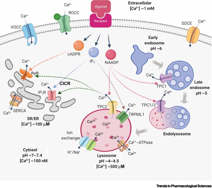
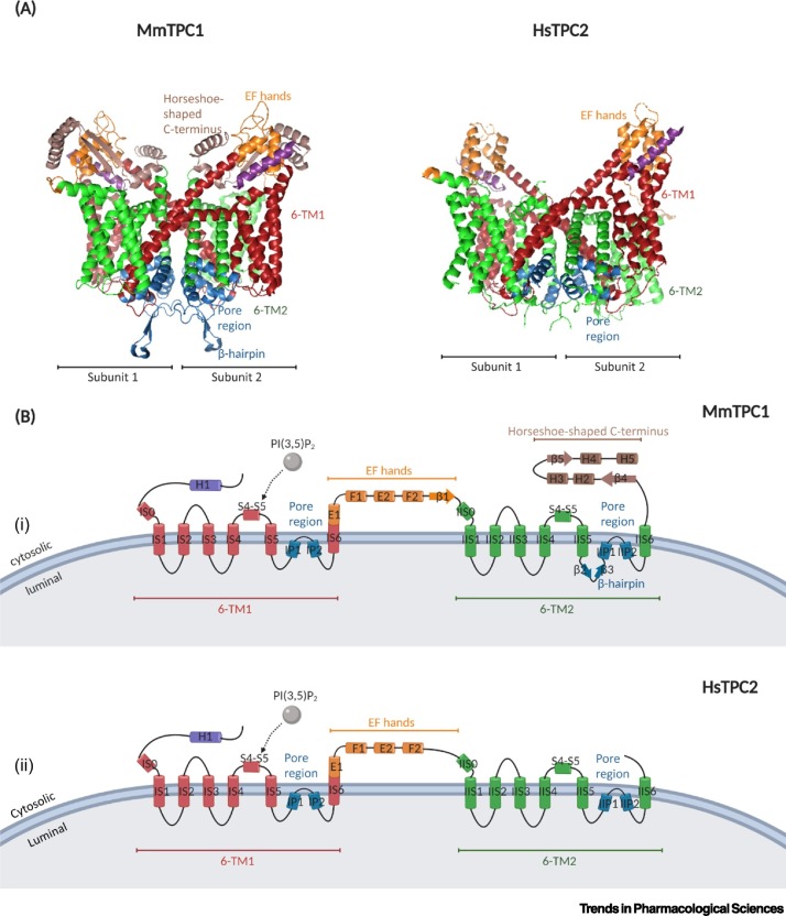
Two-pore channels (TPCs) are cation-permeable channels located on endolysosomal membranes and important mediators of intracellular Ca2+ signalling. TPCs are involved in various pathophysiological processes, including cell growth and development, metabolism, and cancer progression. Most studies of TPCs have used TPC-/- cell or whole-animal models, or Ned-19, an indirect inhibitor. The TPC activation mechanism remains controversial, which has made it difficult to develop selective modulators. Recent studies of TPC structure and their interactomes are aiding the development of direct pharmacological modulators. This process is still in its infancy, but will facilitate future research and TPC targeting for therapeutical purposes. Here, we review the progress of current research into TPCs, including recent insights into their structures, functional roles, mechanisms of activation, and pharmacological modulators.

Keywords: NAADP; calcium; cell signalling; endolysosomes; interactome; two-pore channels.


Figures

Figure 1
Figure 1
Intracellular Ca2+ Homeostasis and Regulation Mechanisms. Cytosolic Ca2+ is crucial in multiple cellular functions. Since extracellular [Ca2+] is significantly higher than intracellular [Ca2+], Ca2+ is released into the cytosol in several ways. Extracellular Ca2+ is transported across the cell membrane through voltage-operated calcium channels (VOCCs), receptor-operated calcium channels (ROCCs), and storage-operated calcium entry (SOCE). Intracellular Ca2+ stores are activated by second messengers when ligands bind to their plasma membrane receptor; these include inositol 1,4,5-trisphosphate (IP3), cyclic adenosine diphosphate ribose (cADPR), and nicotinic acid adenine dinucleotide phosphate (NAADP). Ca2+ in the endo/sarcoplasmic reticulum (ER/SR) is released through ryanodine receptors (RyRs) and inositol 1,4,5-triphosphate receptors (IP3Rs) triggered by cADPR and IP3, respectively. By contrast, acidic Ca2+ stores, which are also known as endolysosomal Ca2+ stores, are gated by two-pore channels (TPCs) and transient receptor potential, mucolipin subfamily 1 (TRPML1), triggered by NAADP and IP3, respectively. Ca2+ can be pumped into ER/SR by the action of sarcoplasmic/endoplasmic reticulum Ca2+-ATPase (SERCA) against a concentration gradient. Notably, the Ca2+-induced Ca2+ release (CICR) phenomenon in SR/ER can be stimulated by TPC-released Ca2+ from acidic Ca2+ stores or by influx of extracellular Ca2+. In the transition from early endosomes to lysosomes, increasing acidity and [Ca2+] can be detected. Created withBioRender.com.
Figure 2
Figure 2
An Overview of the Structure of Mouse Two-Pore Channel 1 (MmTPC1) and Human TPC2 (HsTPC2). (A) Side view of the 3D structure of MmTPC1 [Protein Data Bank (PDB) ID: 6C96] and HsTPC2 (PDB ID: 6NQ1). Each channel contains two identical subunits. There are two six-transmembrane (6-TM) domains in each subunit (shown in red, named IS1–IS6 for subunit 1; and in green, named IIS1–IIS6 for subunit 2), connected by EF-hand motifs (in orange). MmTPC1 is distinguished from HsTPC2 in having a unique β-hairpin structure at the pore region (in blue) and a horseshoe shaped C terminus (in brown). (B) Topology and domain arrangement of a single subunit in MmTPC1 (i) and HsTPC2 (ii). Residues from both 6-TMs constitute the pore region. The binding site of phosphatidylinositol 3,5-bisphosphate [PI(3,5)P2] is located at the first 6-TM. Adapted from [14,15]. Created withBioRender.com.

References

    1. Parrington J., Tunn R. Ca 2+ signals, NAADP and two-pore channels: role in cellular differentiation. Acta Physiol. 2014;211:285–296. - PubMed
    1. Berridge M.J., et al. Calcium signalling: dynamics, homeostasis and remodelling. Nat. Rev. Mol. Cell Biol. 2003;4:517–529. - PubMed
    1. Lee H.C., Aarhus R. A derivative of NADP mobilizes calcium stores insensitive to inositol trisphosphate and cyclic ADP-ribose. J. Biol. Chem. 1995;270:2152–2157. - PubMed
    1. Calcraft P.J., et al. NAADP mobilizes calcium from acidic organelles through two-pore channels. Nature. 2009;459:596–600. - PMC - PubMed
    1. Galione A. NAADP receptors. Cold Spring Harb. Perspect. Biol. 2019;11:a035071. - PMC - PubMed