. 2020 Jul 15;21(14):4992.
doi: 10.3390/ijms21144992.

LY75 Suppression in Mesenchymal Epithelial Ovarian Cancer Cells Generates a Stable Hybrid EOC Cellular Phenotype, Associated with Enhanced Tumor Initiation, Spreading and Resistance to Treatment in Orthotopic Xenograft Mouse Model

Affiliations

LY75 Suppression in Mesenchymal Epithelial Ovarian Cancer Cells Generates a Stable Hybrid EOC Cellular Phenotype, Associated with Enhanced Tumor Initiation, Spreading and Resistance to Treatment in Orthotopic Xenograft Mouse Model

Sadia Mehdi et al. Int J Mol Sci. .

Abstract

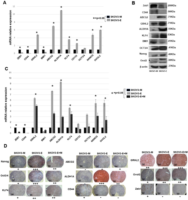
The implications of the epithelial-mesenchymal transition (EMT) mechanisms in the initiation and progression of epithelial ovarian cancer (EOC) remain poorly understood. We have previously shown that suppression of the antigen receptor LY75 directs mesenchymal-epithelial transition (MET) in EOC cell lines with the mesenchymal phenotype, associated with the loss of Wnt/β-catenin signaling activity. In the present study, we used the LY75-mediated modulation of EMT in EOC cells as a model in order to investigate in vivo the specific role of EOC cells, with an epithelial (E), mesenchymal (M) or mixed epithelial plus mesenchymal (E+M) phenotype, in EOC initiation, dissemination and treatment response, following intra-bursal (IB) injections of SKOV3-M (control), SKOV3-E (Ly75KD) and a mixed population of SKOV3-E+M cells, into severe combined immunodeficiency (SCID) mice. We found that the IB-injected SKOV3-E cells displayed considerably higher metastatic potential and resistance to treatment as compared to the SKOV3-M cells, due to the acquisition of a Ly75KD-mediated hybrid phenotype and stemness characteristics. We also confirmed in vivo that the LY75 depletion directs suppression of the Wnt/β-catenin pathway in EOC cells, suggestive of a protective role of this pathway in EOC etiology. Moreover, our data raise concerns regarding the use of LY75-targeted vaccines for dendritic-cell EOC immunotherapy, due to the possible occurrence of undesirable side effects.

Keywords: Ly75; Wnt/β catenin pathway; cancer stem cells (CSCs); epithelial ovarian cancer; epithelial–mesenchymal transition (EMT); hybrid cellular phenotype; orthotopic xenograft mouse model.


Conflict of interest statement

The authors declare no conflict of interest.

Figures

Figure 1
Figure 1
In vivo examination of tumor initiation, spreading, survival and response to treatment in SCID mice IB-injected with SKOV3-M SKOV3-E and SKOV3-E+M cells. (A) Whole body representative bioluminescence images of the emerging tumors and metastatic lesions in SCID mice 59 days, 67 days, 73 days and 130 days post-injection IB of SKOV3-M, SKOV3-E and SKOV3-E+M cells. (B) Survival plots for phase 1 mice IB-injected with SKOV3-M, SKOV3-E and Mix cells. (C) Survival plots for phase 2 mice IB-injected with SKOV3-M and SKOV3-E cells, followed by treatment with carboplatin. Asterisks represent the number of sacrificed mice at each step according to the survival time, as indicated in Table 1; see also Section 4.3.5 for details.
Figure 2
Figure 2
Analysis of the Wnt/B-catenin pathway status in primary tumor tissue samples of phase 1 mice experimental groups IB-injected with SKOV3-M, SKOV3-E or SKOV3-E+M cells. (A) Western blot analysis of β-catenin, APC2 and Axin1 proteins in SKOV3-M (mice 15, 16 and 19), SKOV3-E (mice 21, 22 and 23) and SKOV3-E+M mice (samples 24, 27 and 28). β-actin was used as the loading control. (B) Immunohistochemistry (IHC) analysis of the expression of β-catenin, APC2 and Axin1 and β-catenin sub-cellular localization in tumor tissues extracted from mice injected with the SKOV3-M, SKOV3-E and SKOV3-E+M cells at 40× (scale bar 10 μm). The plus symbols (+ to +++) indicate the level of expression (from lower to higher) of the corresponding protein. (C) Immunofluorescence (IF) analysis of β-catenin, APC2 and Axin1 expression in tumor tissues extracted from mice injected with SKOV3-M and SKOV3-E cells (magnification at 60×). (D) IF analysis of Ly75, β-catenin and β-catenin sub-cellular localization in tumor tissues extracted from mice IB-injected with SKOV3-M and SKOV3-E cells (magnification at 40×).
Figure 3
Figure 3
Protein expression analysis of different EMT markers in SKOV3-M and SKOV3-E cells. (A) Western blot analysis of the protein expression of LY75, N-cadherin, E-cadherin, vimentin and EpCAM in SKOV3-M and SKOV3-E cells. β-actin was used as a loading control. (B) Immunofluorescence analysis of the protein expression of N-cadherin, E-cadherin, vimentin and EpCAM in SKOV3-M and SKOV3-E cells (magnification at 60×).
Figure 4
Figure 4
Protein expression analysis of different EMT markers in tumor tissues extracted from mice IB-injected with SKOV3-M, SKOV3-E and SKOV3-E+M cells. (A) Western blot analysis of the protein expression of LY75, vimentin, N-cadherin and E-cadherin, and EpCAM in tumor tissues extracted from mice IB-injected with SKOV3-M, SKOV3-E and SKOV3-E+M cells. β-actin was used as a loading control. (B) Representative immunohistochemistry (IHC) images of the protein expression of LY75, vimentin, E-cadherin, N-cadherin and EpCAM in tumor tissues extracted from mice injected with the SKOV3-M, SKOV3-E and SKOV3-E+M cells. Magnification at 40× (scale bar 10 μm). The plus symbols (+ to ++++) indicate the level of expression (from lower to higher) of the corresponding protein. The minus (−) symbol means no expression. (C) Immunofluorescence analysis of the protein expression of N-cadherin, vimentin, E-cadherin and EpCAM in tumor tissues extracted from mice IB-injected with SKOV3-M and SKOV3-E cells (magnification at 60×).
Figure 5
Figure 5
Gene (mRNA) and protein expression levels of different CSCs markers and hybrid state stability factors in SKOV3-M and SKOV3-E cells, and in tumor tissues extracted from mice IB-injected with SKOV3-M, SKOV3-E and SKOV3-E+M cells. (A) Quantitative PCR (qPCR) analysis of the mRNA expression levels of some CSCs markers (CD44, BMI1, ABCG2, ALDH1A, KLF4, CD133, OCT3/4 and NANOG) and genes implicated in the control of the hybrid cellular state (ZEB1, GRHL2 and OVOL2), in SKOV3-M and SKOV3-E cells. (B) Western blot analysis of the protein expression levels of some CSCs markers (CD44, ABCG2, ALDH1A, KLF4, BMI1, OCT3/4, Nanog) and genes implicated in the control of the hybrid cellular state (ZEB1, GRHL2 and OVOL2), in SKOV3-M and SKOV3-E cells. (C) qPCR analysis of the mRNA expression levels of some CSCs markers (CD44, BMI1, ABCG2, ALDH1A, KLF4, CD133, OCT3/4 and NANOG) and genes implicated in the control of the hybrid cellular state (ZEB1, GRHL2 and OVOL2), in tumor tissues extracted from mice IB-injected with SKOV3-M, SKOV3-E and SKOV3-E+M cells. Data are presented as mean ± SD. *, p < 0.05; (Student’s t test). (D) Representative IHC images of Nanog, Oct3/4, Klf4, ABCG2, ALDH1A, CD44, GRHL2, OVOL2 and EpCAM expression in tumor tissues extracted from mice injected with the SKOV3-M, SKOV3-E and SKOV3-E+M cells. Magnification at 40× (scale bar 10 μm). The plus symbols (+ to +++) indicate the level of expression (from lower to higher) of the corresponding protein. The minus (−) symbol means no expression.

Similar articles

Cited by

References

    1. Siegel R.L., Miller K.D., Jemal A. Cancer statistics, 2019. Ca A Cancer J. Clin. 2019;69:7–34. doi: 10.3322/caac.21551. - DOI - PubMed
    1. Coleman R.L., Monk B.J., Sood A.K., Herzog T.J. Latest research and treatment of advanced-stage epithelial ovarian cancer. Nat. Rev. Clin. Oncol. 2013;10:211–224. doi: 10.1038/nrclinonc.2013.5. - DOI - PMC - PubMed
    1. Yao D., Dai C., Peng S. Mechanism of the mesenchymal-epithelial transition and its relationship with metastatic tumor formation. Mol. Cancer. Res. 2011;9:1608–1620. doi: 10.1158/1541-7786.MCR-10-0568. - DOI - PubMed
    1. Suhail Y., Cain M.P., Vanaja K., Kurywchak P.A., Levchenko A., Kalluri R. Systems Biology of Cancer Metastasis. Cell Syst. 2019;9:109–127. doi: 10.1016/j.cels.2019.07.003. - DOI - PMC - PubMed
    1. Thiery J.P. Epithelial-mesenchymal transitions in tumour progression. Nat. Rev. Cancer. 2002;2:442–454. doi: 10.1038/nrc822. - DOI - PubMed

MeSH terms