Review
. 2020 Sep 8:74:497-520.
doi: 10.1146/annurev-micro-020518-115638. Epub 2020 Jul 17.

Polymorphic Toxins and Their Immunity Proteins: Diversity, Evolution, and Mechanisms of Delivery

Affiliations
Review

Polymorphic Toxins and Their Immunity Proteins: Diversity, Evolution, and Mechanisms of Delivery

Zachary C Ruhe et al. Annu Rev Microbiol. .

Abstract

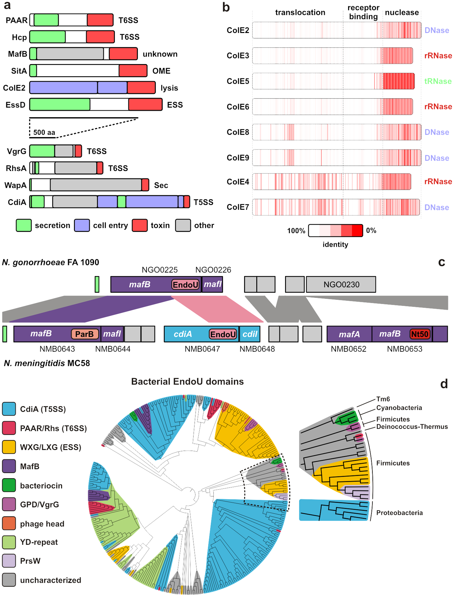
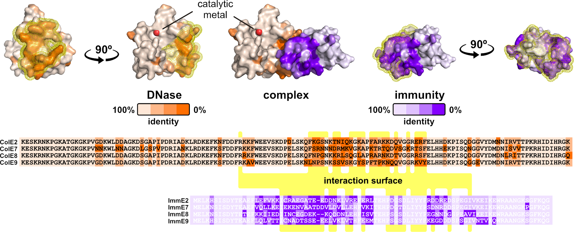
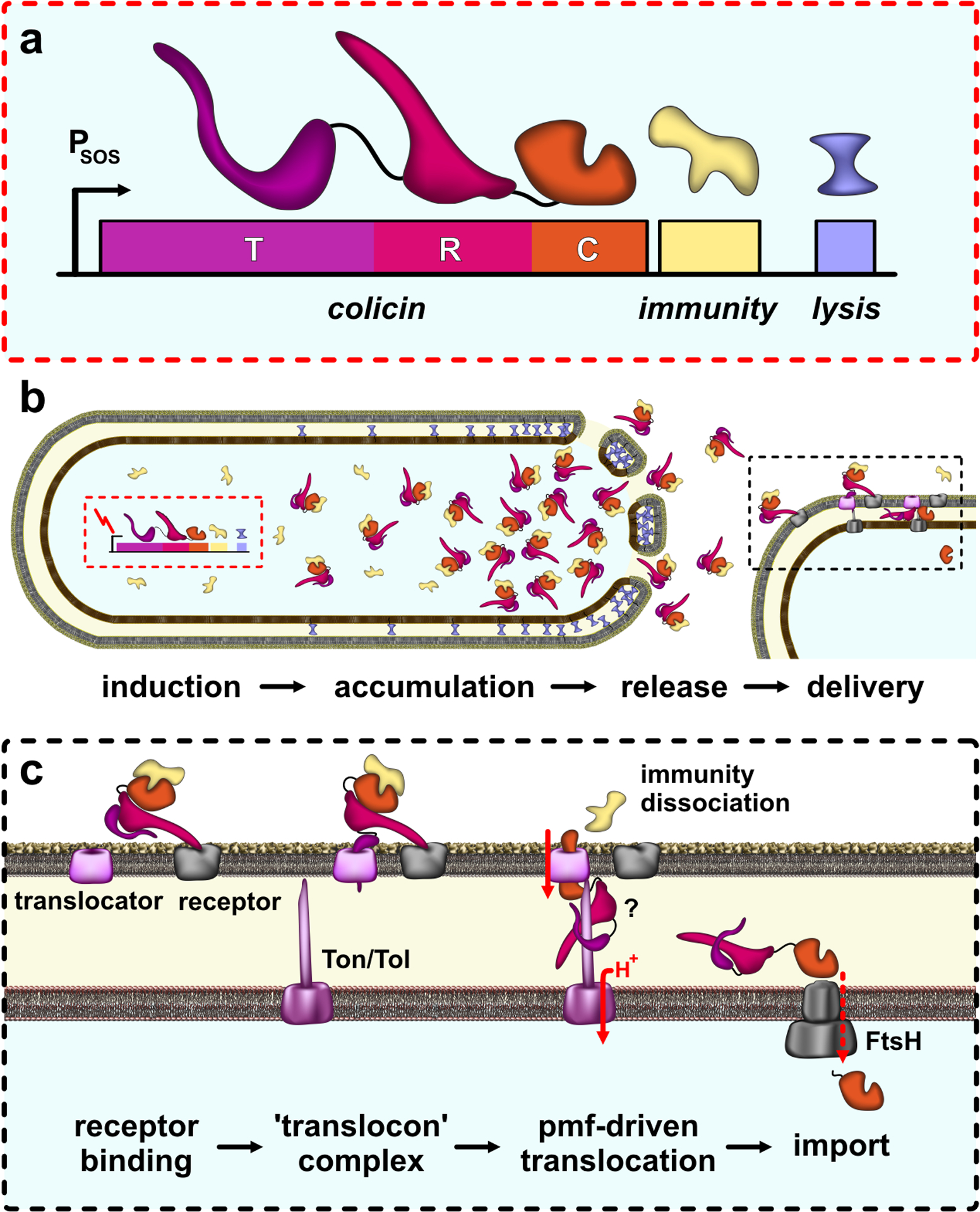
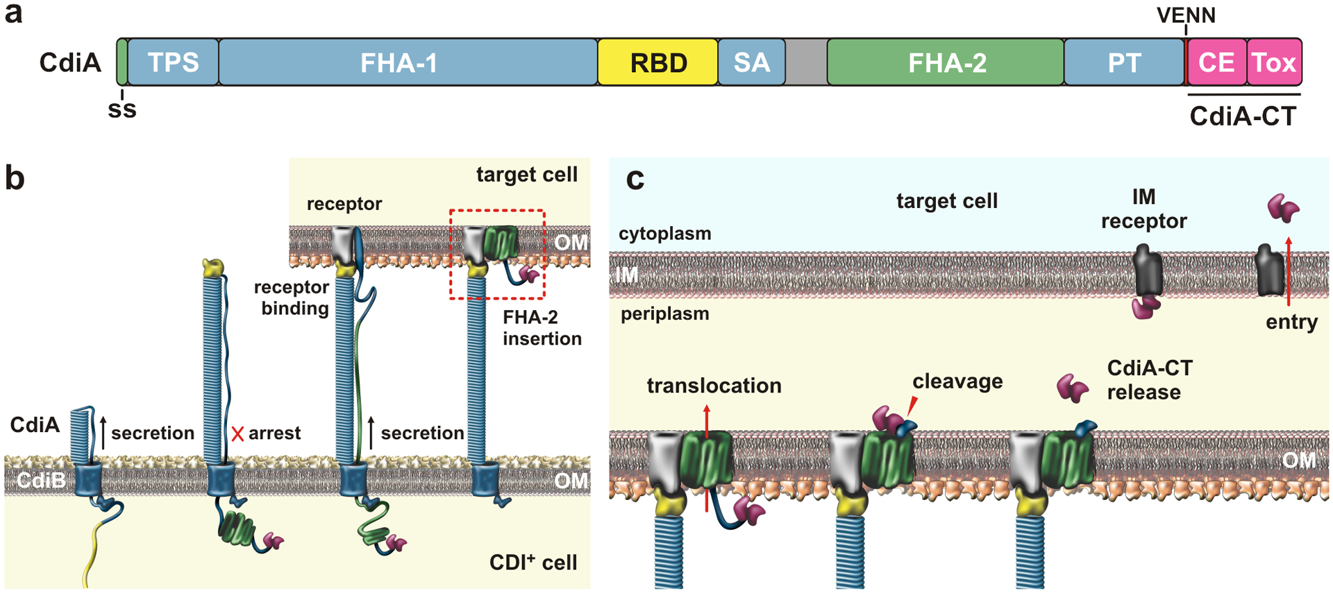
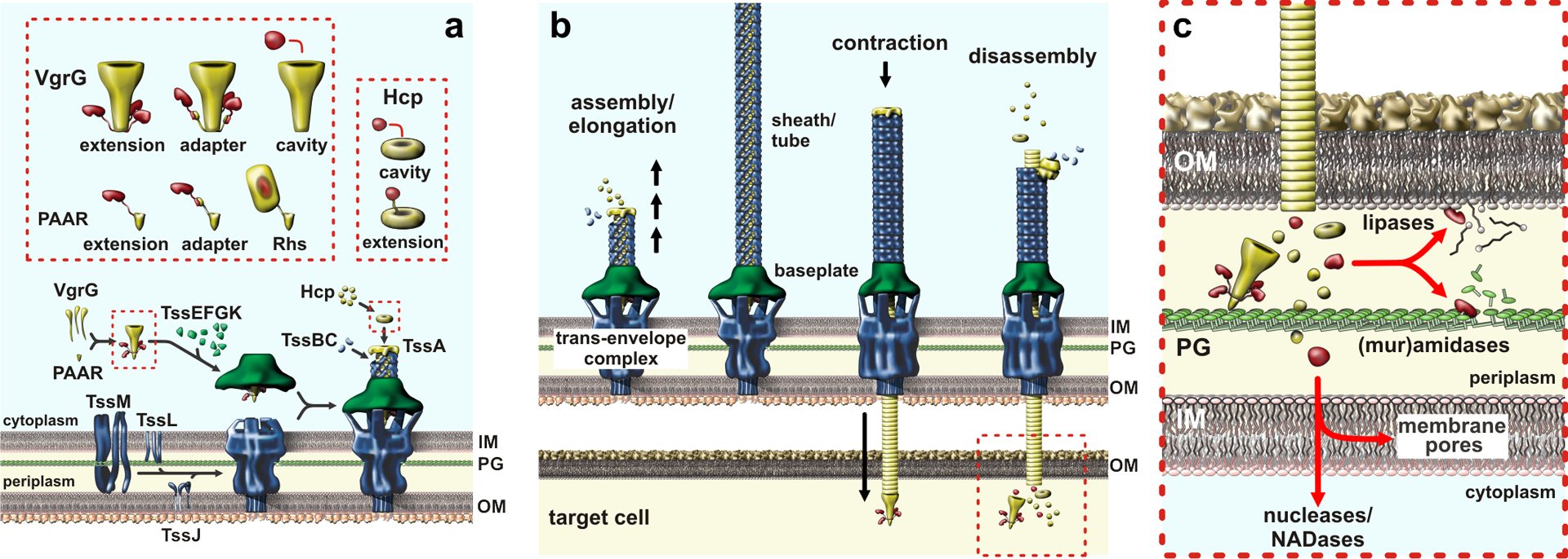
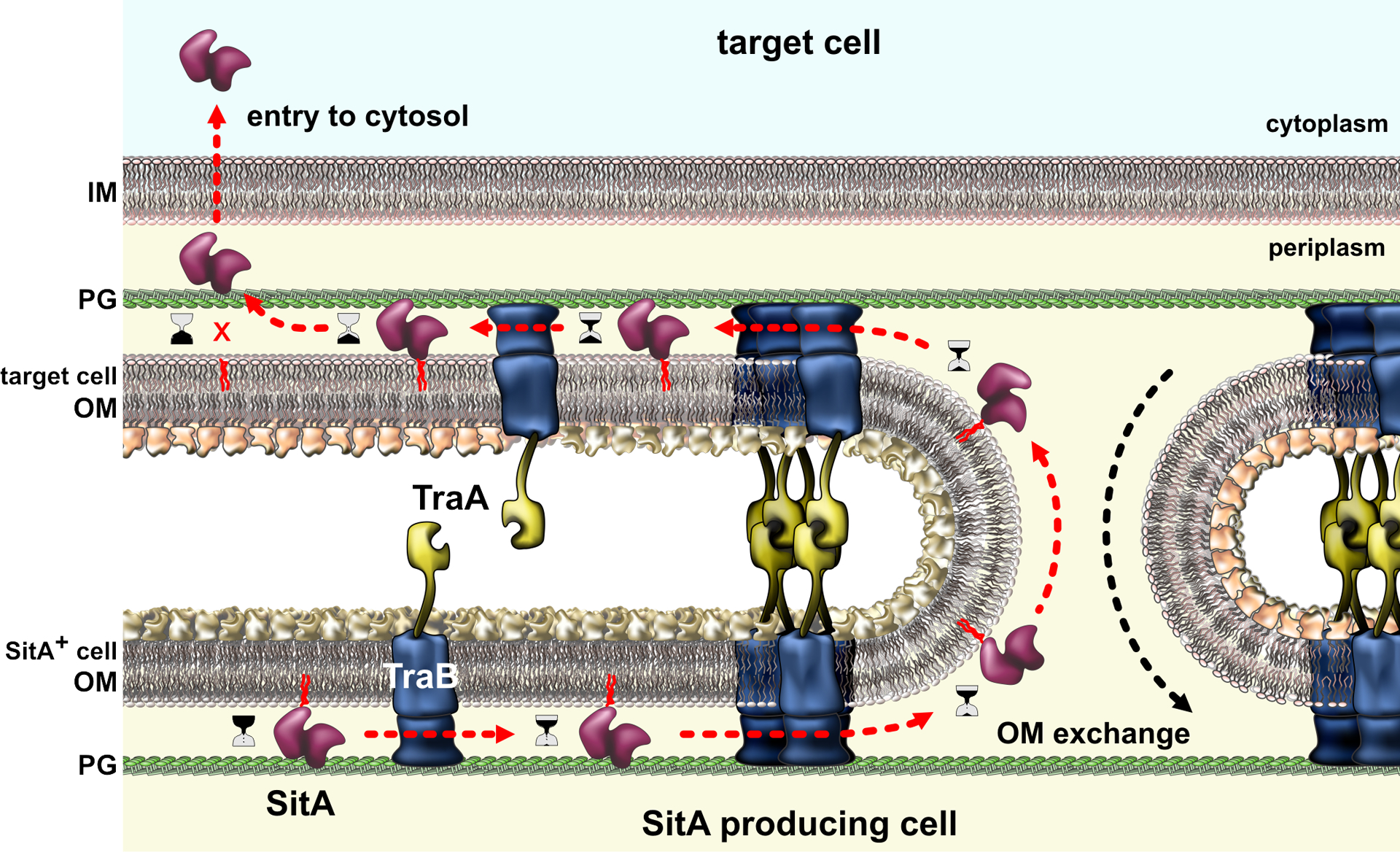
All bacteria must compete for growth niches and other limited environmental resources. These existential battles are waged at several levels, but one common strategy entails the transfer of growth-inhibitory protein toxins between competing cells. These antibacterial effectors are invariably encoded with immunity proteins that protect cells from intoxication by neighboring siblings. Several effector classes have been described, each designed to breach the cell envelope of target bacteria. Although effector architectures and export pathways tend to be clade specific, phylogenetically distant species often deploy closely related toxin domains. Thus, diverse competition systems are linked through a common reservoir of toxin-immunity pairs that is shared via horizontal gene transfer. These toxin-immunity protein pairs are extraordinarily diverse in sequence, and this polymorphism underpins an important mechanism of self/nonself discrimination in bacteria. This review focuses on the structures, functions, and delivery mechanisms of polymorphic toxin effectors that mediate bacterial competition.

Keywords: CDI; ESS; Esx-like secretion system; MafB; OME; T6SS; colicins; contact-dependent growth inhibition; outer membrane exchange; type VI secretion system.


Figures

Figure 1.
Figure 1.. Polymorphic effector families.
a) Polymorphic effector classes that function in bacterial competition. b) E-type colicin domain structure and homology. c) Alignment of maf genomic islands from N. gonorrhoeae FA 1090 and N. meningitidis MC58. Ordered locus identifiers are given and sequences encoding predicted ParB, EndoU and Ntox50 (Nt50) toxin domains are indicated. d) Distribution of EndoU (PF14436) RNase domains across effector classes. Abbreviations: ESS, Esx-like secretion system; Hcp, hemolysin-coregulated protein; OME, outer membrane exchange; T5SS, type V secretion system; T6SS, type VI secretion system.
Figure 2.
Figure 2.. Diversification of toxin-immunity pairs.
Sequence variation is mapped onto the crystal structure of the ColE2 DNase domain bound to its immunity protein. The interaction surface is highlighted with yellow mesh, and residues making direct contact are indicated with yellow in the alignments. The DNase catalytic center is indicated by the divalent metal ion.
Figure 3.
Figure 3.. Colicins.
a) Colicin operon organization. The bacteriocin release protein is encoded by the lysis gene, which is separated from the upstream colicin and immunity cistrons by a transcription terminator. b) Colicin release. Induction of colicin expression allows read-through of the terminator, producing release proteins that disrupt the cell envelope to release colicin-immunity complexes. c) Colicin delivery. Colicins use R domain to recognize OMP receptors on target bacteria, then recruit a second OMP translocator using the T domain. Translocation is energized by the pmf through Tol or Ton systems. Nuclease domains require FtsH for import to the cytosol.
Figure 4.
Figure 4.. Contact-dependent growth inhibition (CDI).
a) CdiA domain architecture. The CdiA-CT is composed of cytoplasm entry (CE) and toxin domains; Abbreviations: ss, signal sequence; TPS, two-partner secretion transport domain; RBD, receptor-binding domain; SA, secretion arrest domain; PT, pretoxin domain; and CE, cytoplasm entry domain. b) CdiA biogenesis. CdiB guides CdiA export across the outer membrane (OM). The C-terminal half of CdiA remains sequestered in the periplasm due to a programmed secretion arrest. CdiA export resumes upon binding receptor, and the FHA-2 domain integrates into the target-cell OM to form a toxin translocation conduit. c) Toxin delivery. Once transferred into the periplasm, the CdiA-CT is released for a second translocation step across the inner membrane (IM). The final step is mediated by cytoplasm entry domains, which interact with membrane proteins and translocate the C-terminal toxin domain in a pmf-dependent manner. Abbreviations: CE, cytoplasm entry domain; CT, C-terminal region; IM, inner membrane; OM, outer membrane; PT, pretoxin domain; RBD, receptor-binding domain; SA, secretion arrest domain; ss, signal sequence; Tox, toxin domain; TPS, two-partner secretion transport domain.
Figure 5.
Figure 5.. Type VI secretion systems.
a) T6SS sub-complex assembly and effector packaging onto VgrG and Hcp. b) Sheath-tube assembly initiates at the baseplate and extends across the width of the cell. The sheath rapidly contracts, expelling the Hcp tube through the baseplate and trans-envelope complexes. The PAAR-tipped VgrG protein penetrates the OM of target bacteria and releases effector proteins. c) T6SS effector proteins activities. Nucleases and NADases are translocated into the cytoplasm through poorly understood pathways. Abbreviations: Hcp, hemolysin-coregulated protein; IM, inner membrane; NADase, NAD(P)+ glycohydrolase; OM, outer membrane; PG, peptidoglycan; Rhs, rearrangement hotspot; T6SS, type VI secretion system; VgrG, valine-glycine repeat G protein.
Figure 6.
Figure 6.. Outer-membrane exchange.
Myxobacteria with compatible TraA proteins engage in homotypic interactions that bring the cells into close approximation for outer-membrane fusion. OM lipids and proteins are freely exchanged between the cells, but TraA itself is not transferred due to interactions with TraB, which is tethered to the peptidoglycan (PG) cell wall. Polymorphic SitA effectors are among the many lipoproteins transferred during OME. SitA nucleases ultimately disengage from the OM and enter the cytoplasm through an uncharacterized mechanism. Abbreviations: IM, inner membrane; OM, outer membrane; PG, peptidoglycan.

References

    1. Ahmad S, Wang B, Walker MD, Tran HR, Stogios PJ, et al. 2019. An interbacterial toxin inhibits target cell growth by synthesizing (p)ppApp. Nature 575:674–8 - PMC - PubMed
    1. Aoki SK, Diner EJ, de Roodenbeke CT, Burgess BR, Poole SJ, et al. 2010. A widespread family of polymorphic contact-dependent toxin delivery systems in bacteria. Nature 468:439–42 - PMC - PubMed
    1. Aoki SK, Malinverni JC, Jacoby K, Thomas B, Pamma R, et al. 2008. Contact-dependent growth inhibition requires the essential outer membrane protein BamA (YaeT) as the receptor and the inner membrane transport protein AcrB. Mol. Microbiol 70:323–40 - PMC - PubMed
    1. Aoki SK, Pamma R, Hernday AD, Bickham JE, Braaten BA, Low DA. 2005. Contact-dependent inhibition of growth in Escherichia coli. Science 309:1245–8 - PubMed
    1. Aoki SK, Webb JS, Braaten BA, Low DA. 2009. Contact-dependent growth inhibition causes reversible metabolic downregulation in Escherichia coli. J. Bacteriol 191:1777–86 - PMC - PubMed

Publication types

MeSH terms