Case Reports
. 2020 Jul 17;8(1):111.
doi: 10.1186/s40478-020-00992-9.

Global activation of oncogenic pathways underlies therapy resistance in diffuse midline glioma

Affiliations
Case Reports

Global activation of oncogenic pathways underlies therapy resistance in diffuse midline glioma

M-M Georgescu et al. Acta Neuropathol Commun. .

Abstract

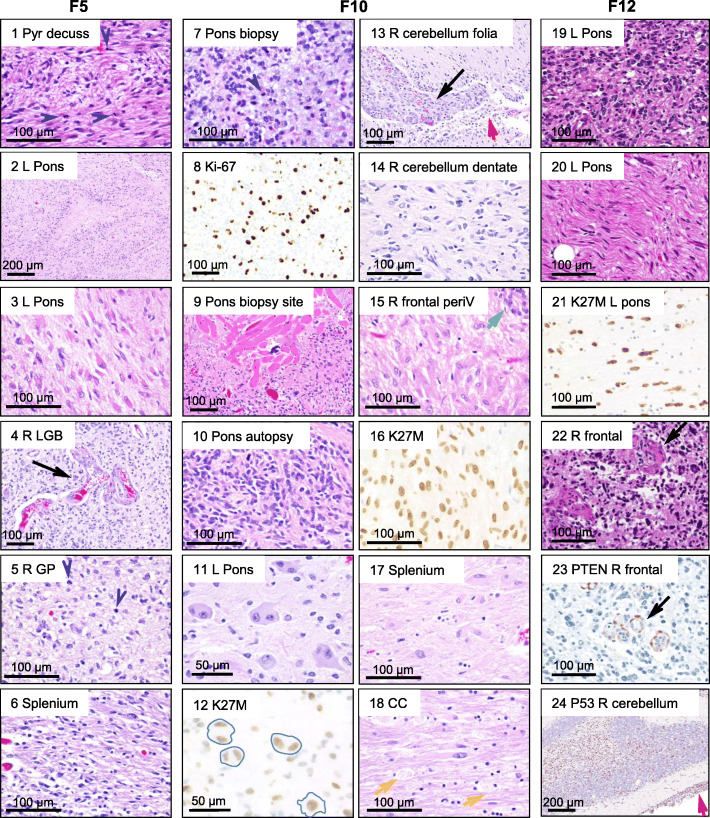
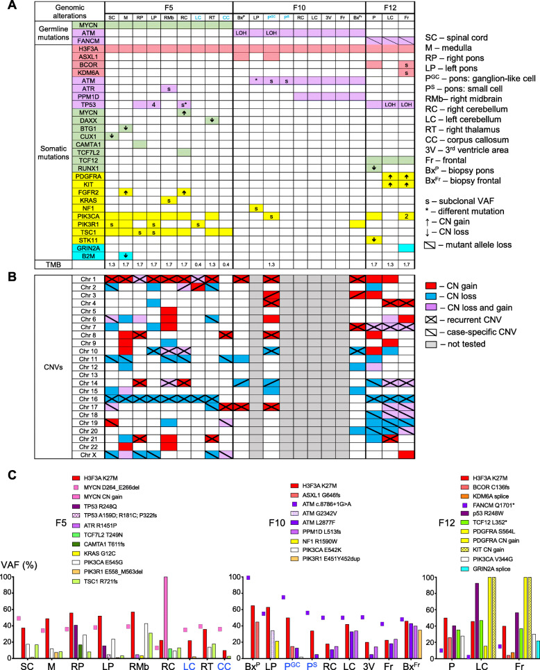
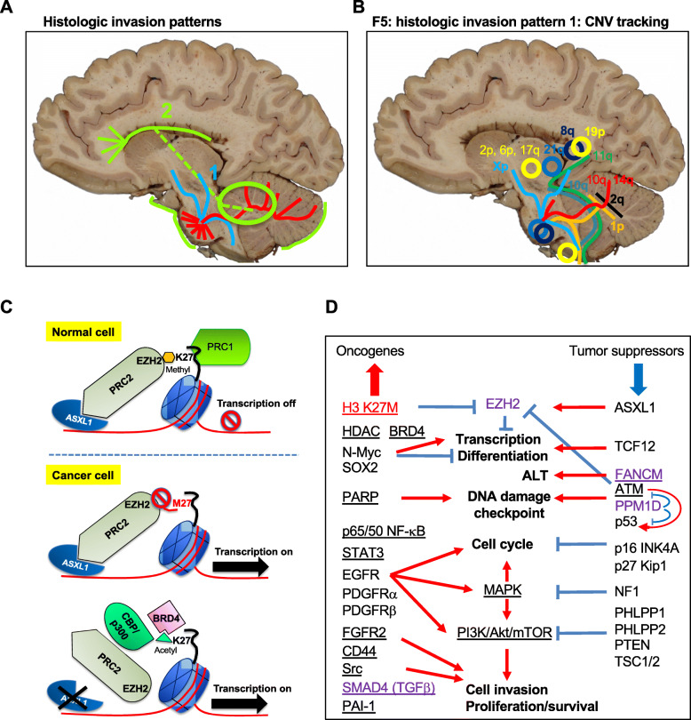
Diffuse midline gliomas (DMGs) are aggressive pediatric brain tumors with dismal prognosis due to therapy-resistant tumor growth and invasion. We performed the first integrated histologic/genomic/proteomic analysis of 21 foci from three pontine DMG cases with supratentorial dissemination. Histone H3.3-K27M was the driver mutation, usually at high variant allele fraction due to recurrent chromosome 1q copy number gain, in combination with germline variants in ATM, FANCM and MYCN genes. Both previously reported and novel recurrent copy number variations and somatic pathogenic mutations in chromatin remodeling, DNA damage response and PI3K/MAPK growth pathways were variably detected, either in multiple or isolated foci. Proteomic analysis showed global upregulation of histone H3, lack of H3-K27 trimethylation, and further impairment of polycomb repressive complex 2 by ASXL1 downregulation. Activation of oncogenic pathways resulted from combined upregulation of N-MYC, SOX2, p65/p50 NF-κB and STAT3 transcription factors, EGFR, FGFR2, PDGFRα/β receptor tyrosine kinases, and downregulation of PHLPP1/2, PTEN and p16/INK4A tumor suppressors. Upregulation of SMAD4, PAI-1, CD44, and c-SRC in multiple foci most likely contributed to invasiveness. This integrated comprehensive analysis revealed a complex spatiotemporal evolution in diffuse intrisic pontine glioma, recommending pontine and cerebellar biopsies for accurate populational genetic characterization, and delineated common signaling pathways and potential therapeutic targets. It also revealed an unsuspected activation of a multitude of oncogenic pathways, including cancer cell reprogramming, explaining the resistance of DMG to current therapies.

Keywords: Diffuse midline glioma, Autopsy, Next generation sequencing (NGS), Copy number variation (CNV), Proteomics.


Conflict of interest statement

The authors declare that they have no competing interests.

Figures

Fig. 1
Fig. 1
Histologic patterns of tumor cell invasion in DIPG. a. Brain gross appearance. Red and green arrows indicate the primary pontine and the secondary cerebellar foci, respectively. R, right; L, left. b. Sections of indicated brain structures with gross visible tumor foci. Black and blue arrows indicate liquefying necrosis and fleshy tumor, respectively. c. Semiquantitative analysis of histological tumor involvement of the specified color-coded structures. MVP, microvascular proliferation; 4 V, 4th ventricle; PF, posterior fossa; CN, cranial nerve; 3 V, 3rd ventricle; CC, corpus callosum; Temp, temporal; Occip, occipital; WM, white matter; Pyr, pyramidal tracts; IC, internal capsule. The panels and graphs from (b) and (c), respectively, correspond to the brains from (a)
Fig. 2
Fig. 2
Morphologic spectrum of the 3 DIPGs across infratentorial and supratentorial foci. H&E and IHC with Ki-67, histone H3-K7M, PTEN and p53 antibodies of selected sections indicated by numbers and anatomic locations. The columns correspond to each case, with two central columns shown for F10. L, left; R, right; Pyr decuss, pyramidal tract decussation; LGB, lateral geniculate body; GP, globus pallidus; periV, periventricular; CC, corpus callosum. Blue arrowheads and black, red, green and yellow arrows indicate mitotic figures, microvascular proliferation, leptomeningeal spread, subventricular ependymal cell proliferation, and foamy macrophages, respectively. Among other features, note various neoplastic cell morphologies, supratentorial involvement for all tumors, brisk mitotic activity in multiple F5 foci and in the F10 biopsy, viable striated muscle fibers in F10, most likely at prior biopsy site (panel 9), labeling with H3-K27M antibody of F10 bi-nucleated ganglion cells (blue contour in panel 12), and PTEN expression detected in endothelial cells but not in tumor cells, in F12
Fig. 3
Fig. 3
Spatio-temporal genomic profiling. a. Pathway color-coded representation of gene mutations and selected CNVs: chromatin remodeling – pink; DDR – purple; transcription factors and modulators – green; growth factor receptors and mediators – yellow; other – blue. White boxes lack mutations, and numbers in boxes indicate the number of mutant variants for the respective gene. FFPE microdissected tumor foci are showed in blue font and show decreased TBM for the F5 samples, indicating limited tumor material available for NGS that most likely lowered the mutation and CNV detection rate in these samples. b. Color-coded CNV analysis. c. VAF graphic representation: germline mutations as dots, somatic mutations as bars. FFPE-microdissected samples are shown in blue font and show decreased VAF for the F5 samples
Fig. 4
Fig. 4
Proteomic profiling of DIPG foci. a-b. WB analysis with indicated antibodies of total protein lysates (50 μg proteins) from tumor (T) and normal (N) fresh frozen autopsy samples. Primary pontine (P) foci are indicated in red font and secondary cerebellar (C) and frontal (Fr) foci, in green font. Sample laterality: R, right; L, left. Supplemental Fig. S6 shows the positive control for H3 K27Me3 antibody from an adult glioblastoma autopsy case with EZH2 overexpression. Note lack of H3 K27 methylation in the presence of EZH2 overexpression for F12 right frontal (RFr) tumor focus. c. Heat map of semiquantitative WB analysis, as shown quantified and normalized to loading controls in Supplemental Fig. S6. Shades of red and blue indicate expression level increase or decrease, respectively, as compared to normal controls
Fig. 5
Fig. 5
Models of invasion, pathway activation and therapeutic targeting in DIPG. a. Histologic invasion pathways include the common ponto-cerebellar dissemination pathway, shown in red, and the more specific centrifugal and CSF/leptomeningeal pathways shown in blue and green, respectively. The green circle indicates the cerebellar tumor as the most common origin of supratentorial CSF seeding. b. Spatial color-coded CNV-tracking of invasive neoplastic populations within the F5 tumor that showed histologic centrifugal migration pattern. Contiguous neoplastic populations are indicated with line tracings, and isolated foci sharing the same CNV composition, with circles. Cerebellar bars indicate bilateral spread of the respective population. The corresponding color-coded CNVs are indicated in bold for gains, and regular font, for losses. c. Schematic model of epigenetic changes targeting histone H3 K27 residue. The histone H2A/H2B/H3/H4 nucleosome is represented as a blue barrel, the DNA, as a red thread, and the N-terminal histone H3 “tail”, as a black curved line. The methyl and acetyl groups are shown in orange and green, respectively. The stop sign shows blockage of enzymatic or transcriptional activity. d. Pathway activation and drug targeting model in DIPG. The oncogenic H3 K27M driver protein and mediators with dual tumor suppressor or oncogenic roles depending on context are shown in red and purple, respectively. Otherwise, the oncogenic and the tumor suppressor proteins are shown on left and right, respectively. The pathways are shown in bold font, and red and blue indicate activation or suppression, respectively. The candidate targets for therapy are underlined

References

    1. Louis DN, Ohgaki H, Wiestler OD, Caveneee WK. WHO classification of Tumors of the central nervous system. Lyon: IARC; 2016. - PubMed
    1. Chiang JC, Ellison DW. Molecular pathology of paediatric central nervous system tumours. J Pathol. 2017;241:159–172. - PubMed
    1. Benitez-Ribas D, Cabezon R, Florez-Grau G, Molero MC, Puerta P, Guillen A, et al. Immune response generated with the Administration of Autologous Dendritic Cells Pulsed with an Allogenic Tumoral cell-lines lysate in patients with newly diagnosed diffuse intrinsic Pontine Glioma. Front Oncol. 2018;8:127. - PMC - PubMed
    1. Chheda ZS, Kohanbash G, Okada K, Jahan N, Sidney J, Pecoraro M, et al. Novel and shared neoantigen derived from histone 3 variant H3.3K27M mutation for glioma T cell therapy. J Exp Med. 2018;215:141–157. - PMC - PubMed
    1. Mathew RK, Rutka JT. Diffuse intrinsic Pontine Glioma : clinical features, molecular genetics, and novel targeted therapeutics. J Korean Neurosurg Soc. 2018;61:343–351. - PMC - PubMed

Publication types