Review
. 2020 Jul 17;147(14):dev190629.
doi: 10.1242/dev.190629.

Mechanisms of human embryo development: from cell fate to tissue shape and back

Affiliations
Review

Mechanisms of human embryo development: from cell fate to tissue shape and back

Marta N Shahbazi. Development. .

Abstract

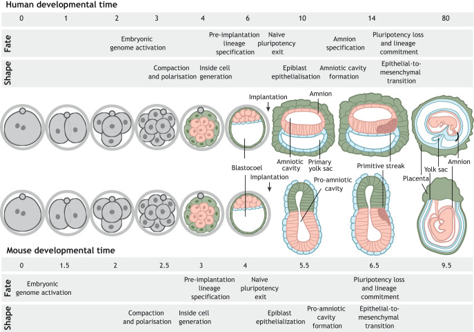
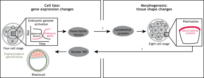
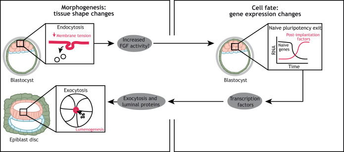
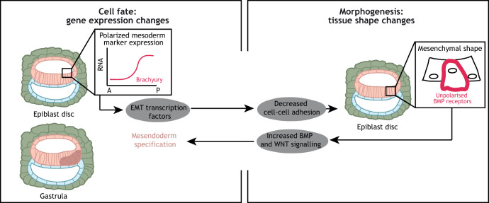
Gene regulatory networks and tissue morphogenetic events drive the emergence of shape and function: the pillars of embryo development. Although model systems offer a window into the molecular biology of cell fate and tissue shape, mechanistic studies of our own development have so far been technically and ethically challenging. However, recent technical developments provide the tools to describe, manipulate and mimic human embryos in a dish, thus opening a new avenue to exploring human development. Here, I discuss the evidence that supports a role for the crosstalk between cell fate and tissue shape during early human embryogenesis. This is a critical developmental period, when the body plan is laid out and many pregnancies fail. Dissecting the basic mechanisms that coordinate cell fate and tissue shape will generate an integrated understanding of early embryogenesis and new strategies for therapeutic intervention in early pregnancy loss.

Keywords: Aneuploidy; Cell fate; Embryonic stem cells; Human embryo; Morphogenesis; Polarisation.


Conflict of interest statement

Competing interestsThe authors declare no competing or financial interests.

Figures

Fig. 1.
Fig. 1.
Overview of human and mouse embryo development. Upon fertilisation, mouse and human embryos undergo a series of cleavage divisions. The embryonic genome becomes activated by the two-cell stage in mouse embryos and at the four/eight-cell stage transition in human embryos. It is followed by compaction and polarisation, which occur at the eight-cell stage in mouse embryos, and between the eight- to 16-cell stage in human embryos. Formation of a hollow cavity, the blastocoel, denotes the formation of the blastocyst, which, by embryonic day E4 (mouse) and E6 (human), is composed of three main tissues: epiblast, hypoblast and trophectoderm. Upon implantation (E5 in mouse and E7 in human), embryos undergo a global morphological transformation. The embryonic epiblast loses its naïve pluripotent character, becomes epithelial and forms the pro-amniotic cavity (mouse), which spans both the epiblast and the trophectoderm-derived extra-embryonic ectoderm, and the amniotic cavity (human). A key difference between mouse and human embryos relates to the formation of the amnion. Whereas in mouse embryos amnion formation takes place during gastrulation, in human embryos a subset of epiblast cells differentiates to form the squamous amniotic epithelium during early post-implantation (E10). As a result, the human epiblast acquires a disc shape and the mouse epiblast a cylindrical morphology. On embryonic days 6.5 (mouse) and 14 (human), gastrulation is initiated in the posterior epiblast. Cells undergo an epithelial-to-mesenchymal transition, lose their pluripotent character and commit to one of the germ layers. Epiblast-derived tissues are shown in pink, hypoblast-derived tissues are shown in blue and trophectoderm-derived tissues are shown in green.
Fig. 2.
Fig. 2.
Proposed cell fate-tissue shape crosstalk during human pre-implantation development. The onset of apicobasal polarisation at the eight-cell stage may be controlled by the expression of embryonic genes, potentially encoding transcription factors. As recently shown in mouse embryos, embryonic genome activation induces the expression of regulators of the actin cytoskeleton. This leads to a remodelling of the actin network that is necessary for the clustering of apical polarity proteins, and culminates in the formation of the mature apical domain. In turn, the acquisition of apicobasal polarisation may lead to nuclear YAP localisation, as seen in the mouse, and expression of trophectoderm-specific transcription factors. This model remains to be functionally validated in human embryos. Question marks denote molecular connections that have not been validated in human embryos.
Fig. 3.
Fig. 3.
Proposed cell fate-tissue shape crosstalk during human implantation development. Upon implantation, epiblast cells exit from the naïve pluripotent state and initiate the expression of post-implantation factors. Studies in mouse ESCs have shown that this is controlled by a decrease in membrane tension, which promotes endocytosis and, as a consequence, increased fibroblast growth factor (FGF) signalling activity, which is required for naïve pluripotency exit. Therefore, membrane tension affects the pluripotent state of a cell. Whether this pathway is active in human embryos remains to be explored. Upon naïve pluripotency exit, genes involved in exocytosis become expressed and initiate the process of lumenogenesis to form the amniotic cavity. Exocytic vesicles provide apical membrane and luminal proteins, which are necessary to build a lumen de novo. The transcription factors involved in this process remain to be identified. Question mark denotes a molecular event that has not beem validated in human embryos.
Fig. 4.
Fig. 4.
Proposed cell fate-tissue shape crosstalk during human post-implantation development. BMP and WNT signals secreted by the amnion and the trophectoderm lead to expression of mesoderm markers, such as the transcription factor brachyury, in the posterior epiblast. Brachyury induces expression of epithelial-to-mesenchymal transition (EMT) transcription factors, which cause a decrease in the levels of E-cadherin. As a consequence, cell-cell adhesions weaken and cells lose their epithelial morphology and their polarised organisation, such as the polarised localisation of BMP receptors. The unpolarised BMP receptors are able to interact with BMP ligands, increasing BMP signalling activity and inducing mesendoderm specification.

References

    1. Alarcon V. B. (2010). Cell polarity regulator PARD6B is essential for trophectoderm formation in the preimplantation mouse embryo. Biol Reprod 83, 347-358. 10.1095/biolreprod.110.084400 - DOI - PMC - PubMed
    1. Alexandrova S., Kalkan T., Humphreys P., Riddell A., Scognamiglio R., Trumpp A. and Nichols J. (2016). Selection and dynamics of embryonic stem cell integration into early mouse embryos. Development 143, 24-34. 10.1242/dev.124602 - DOI - PMC - PubMed
    1. Alikani M. (2005). Epithelial cadherin distribution in abnormal human pre-implantation embryos. Hum Reprod 20, 3369-3375. 10.1093/humrep/dei242 - DOI - PubMed
    1. Amita M., Adachi K., Alexenko A. P., Sinha S., Schust D. J., Schulz L. C., Roberts R. M. and Ezashi T. (2013). Complete and unidirectional conversion of human embryonic stem cells to trophoblast by BMP4. Proc Natl Acad Sci U S A 110, E1212-E1221. 10.1073/pnas.1303094110 - DOI - PMC - PubMed
    1. Anani S., Bhat S., Honma-Yamanaka N., Krawchuk D. and Yamanaka Y. (2014). Initiation of Hippo signaling is linked to polarity rather than to cell position in the pre-implantation mouse embryo. Development 141, 2813-2824. 10.1242/dev.107276 - DOI - PubMed

Publication types