. 2020 Jul 17;10(1):11910.
doi: 10.1038/s41598-020-68438-9.

Platelet-primed interactions of coagulation and anticoagulation pathways in flow-dependent thrombus formation

Affiliations

Platelet-primed interactions of coagulation and anticoagulation pathways in flow-dependent thrombus formation

Sanne L N Brouns et al. Sci Rep. .

Abstract

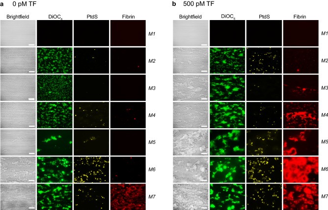
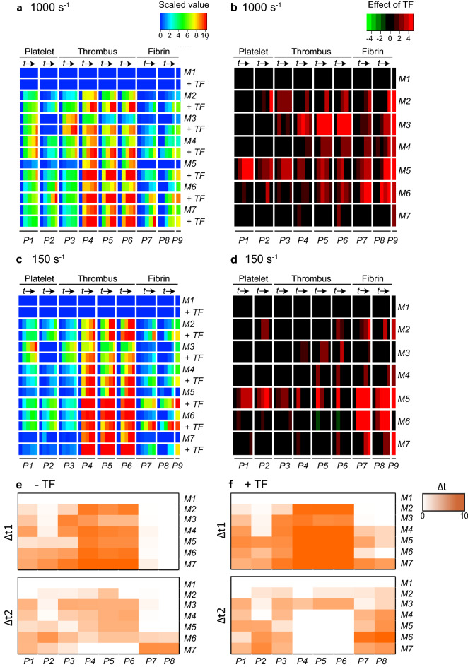
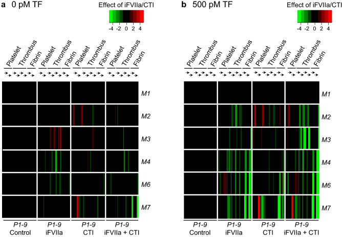
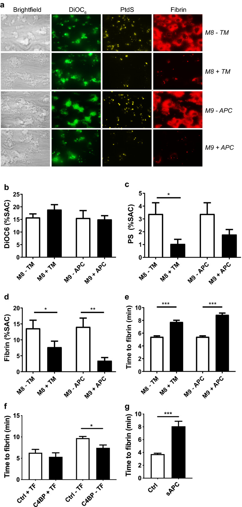
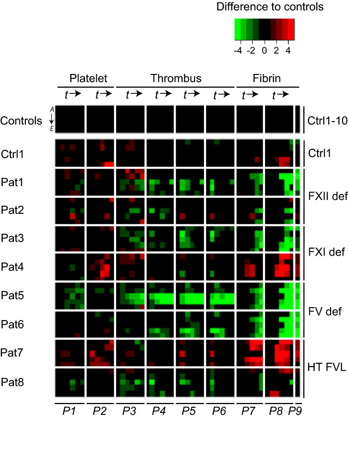
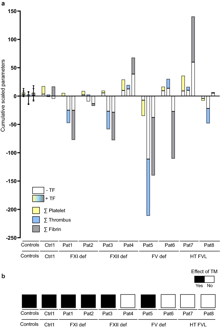
In haemostasis and thrombosis, platelet, coagulation and anticoagulation pathways act together to produce fibrin-containing thrombi. We developed a microspot-based technique, in which we assessed platelet adhesion, platelet activation, thrombus structure and fibrin clot formation in real time using flowing whole blood. Microspots were made from distinct platelet-adhesive surfaces in the absence or presence of tissue factor, thrombomodulin or activated protein C. Kinetics of platelet activation, thrombus structure and fibrin formation were assessed by fluorescence microscopy. This work revealed: (1) a priming role of platelet adhesion in thrombus contraction and subsequent fibrin formation; (2) a surface-independent role of tissue factor, independent of the shear rate; (3) a mechanism of tissue factor-enhanced activation of the intrinsic coagulation pathway; (4) a local, suppressive role of the anticoagulant thrombomodulin/protein C pathway under flow. Multiparameter analysis using blood samples from patients with (anti)coagulation disorders indicated characteristic defects in thrombus formation, in cases of factor V, XI or XII deficiency; and in contrast, thrombogenic effects in patients with factor V-Leiden. Taken together, this integrative phenotyping approach of platelet-fibrin thrombus formation has revealed interaction mechanisms of platelet-primed key haemostatic pathways with alterations in patients with (anti)coagulation defects. It can help as an important functional add-on whole-blood phenotyping.


Conflict of interest statement

J.W.H. is founder and co-owner at FlowChamber b.v. RWF is founder of CambCol Laboratories Ltd. The other authors declare no competing interests.

Figures

Figure 1
Figure 1
Simultaneous analysis of platelet deposition, thrombus phenotype and fibrin formation during whole blood thrombus formation under flow. Citrated whole blood from control subjects (n = 5–10) was supplemented with fluorescent labels to simultaneously detect platelet adhesion (DiOC6), procoagulant, phosphatidylserine (PtdS)-exposing platelets (AF568-annexin A5) and fibrin formation (AF647-fibrinogen). Blood samples were co-perfused with CaCl2/MgCl2 over indicated microspots, at a shear rate of 1,000 s-1. Microspot coding: M1, blocking buffer BSA; M2, rhodocytin + VWF; M3, laminin + VWF; M4, collagen-III; M5, collagen-I low; M6, collagen-I high; M7, GFOGER-GPO + VWF-BP; co-coating with 500 pM TF as indicated. (A) Representative bright-field and fluorescence images, taken from thrombi on microspots without TF (6 min). (B) Idem, from thrombi on microspots co-coated with 500 pM TF (6 min). Bars = 20 µm.
Figure 2
Figure 2
Surface-dependent enhancement by tissue factor of kinetics of platelet deposition, thrombus and fibrin formation. Whole blood with fluorescent labels was flowed over microspots M1–7 with or without co-coated TF (500 pM), as for Fig. 2. Multicolour microscopic images were captured every 2 min (t = 0, 2, 4, 6, 8 min, →), and analysed for parameters P1–9 (for coding, see Table 1). (A,B) Blood flow at high shear rate (1,000 s-1). (C,D) Blood flow at low shear rate (150 s-1). (A,C) Heatmaps of univariate scaled (0–10) values per parameter (P1–9) across surfaces with(out) co-coated TF (500 pM). (B,D) Subtraction heatmaps indicating relevant TF effects per parameter, filtered for changes outside range of mean ± SD. Means of flow runs, n = 5–10 subjects. Scaling was at 0–10 across all surfaces. (E,F) Differential increase in parameters P1–9 (1,000 s-1) over intervals Δt1 = (0–4) min and Δt2 = (4–8) min per microspot without (E) or with (F) TF. Colour code indicates parameter increase (scaled 0–10 across surfaces, 8 min). Full data are given in Data File 1.
Figure 3
Figure 3
Additive contribution of extrinsic and intrinsic coagulation pathways to kinetics of whole blood thrombus formation. Blood from control subjects was collected into citrate with or without CTI (40 μg/mL); where indicated, samples were supplemented with iFVIIa (1 μM). Whole blood perfusion (1,000 s-1) was over microspots M1–7 (± co-coated TF, 500 pM). Analysis of parameters P1–9 over time ( →) was carried out, as for Fig. 2. Four conditions (± iFVIIa ± CTI) were compared per subject (n = 4–6). Data were scaled (0–10) per parameter (P1–9) across surfaces. (A) Subtraction heatmap representing relevant effects of CTI, iFVIIa or CTI + iFVIIa (no TF). (B) Idem for microspots with TF. Effects were filtered for changes outside the range of means ± SD. Data File 3 provides full data.
Figure 4
Figure 4
Alterations in whole blood thrombus formation by immobilized thrombomodulin or activated protein C (APC). (AE) Collagen-I microspots were co-coated or not with TM (M8) or APC (M9), and subjected to whole-blood flow at 1,000 s-1 (TF in recalcification medium). (A) Representative bright-field and fluorescence microscopic images, as in Fig. 1, taken after 6 min; bars = 20 µm. (BD) Platelet deposition (B), phosphatidylserine (C) and fibrin formation (D) after 6 min of flow. (E), Time to fibrin formation per microspotted surface, demonstrating delaying effects of TM and APC. Means ± SEM. n = 10–11, *p < 0.05. **p < 0.01, ***p < 0.001. (F,G) C4BP or soluble APC (sAPC) were added to whole blood before flow perfusion over collagen-I microspots co-coated or not with TF (M6). Time to fibrin formation (n = 4–7) in the presence of C4BP (F) or sAPC (G).
Figure 5
Figure 5
Altered thrombus and fibrin formation in patients with coagulation factor abnormalities. Blood samples from healthy day control subjects (n = 10) and patients (Pat, n = 8) with indicated coagulation factor abnormalities were investigated for platelet, thrombus, and fibrin formation at a shear rate of 1,000 s-1, with image capturing every 2 min (see Fig. 2). Image parameters (P1–9) were obtained for microspot M6 no TF (rows A) or with TF (rows B); M7 no TF (rows C) or with TF (rows D); M8 with TF (rows E). Data were univariate scaled (0–10) across surfaces per parameter for all flow runs. Differential heatmap of scaled parameters over time per patient in comparison to means of control subjects. Filtering applied for changes outside the range of means ± SD of control subjects. A green colour indicates a relevant decrease, a red colour a relevant increase in comparison to controls. Full data are given in Data File 5.
Figure 6
Figure 6
Integrated changes in thrombus formation in the presence or absence of TF or TM for individual patients with coagulation factor abnormalities. Microspot-based formation of platelet–fibrin thrombi was measured in whole-blood from 10 control subjects and 8 patients with coagulation factor abnormalities, as in Fig. 5. Relevant changes of scaled parameters in comparison to means of control subjects (Ctrl, outside range of means ± SD) were integrated for all time points. Bars show results from all controls (means ± SD), from a representative control (Ctr1) and all individual patients (Pat1 to Pat8). (A) Cumulative relevant changes of scaled parameters of platelet adhesion (P1–2), thrombus signature (P3–6) and fibrin formation (P7–9) across five surfaces. (B) Mean relevant effects of TM on fibrin parameters for control subjects and patients, assessed for M8.

References

    1. Heemskerk JW, Mattheij N, Cosemans JM. Platelet-based coagulation: different populations, different functions. J. Thromb. Haemost. 2013;11:2–11. - PubMed
    1. Alshehri OM, et al. Fibrin activates GPVI in human and mouse platelets. Blood. 2015;126:1601–1608. - PMC - PubMed
    1. Mammadova-Bach E, et al. Platelet glycoprotein VI binds to polymerized fibrin and promotes thrombin generation. Blood. 2015;126:683–691. - PubMed
    1. Podoplelova NA, et al. Coagulation factors bound to procoagulant platelets concentrate in cap structures to promote clotting. Blood. 2016;128:1745–1755. - PubMed
    1. Mackman N, Tilley RE, Key NS. Role of the extrinsic pathway of blood coagulation in hemostasis and thrombosis. Arterioscler. Thromb. Vasc. Biol. 2007;27:1687–1693. - PubMed

Publication types