Case Reports
. 2020 Sep 8;154(4):466-474.
doi: 10.1093/ajcp/aqaa124.

SARS-CoV-2 Infection-Associated Hemophagocytic Lymphohistiocytosis

Affiliations
Case Reports

SARS-CoV-2 Infection-Associated Hemophagocytic Lymphohistiocytosis

Andrey Prilutskiy et al. Am J Clin Pathol. .

Abstract

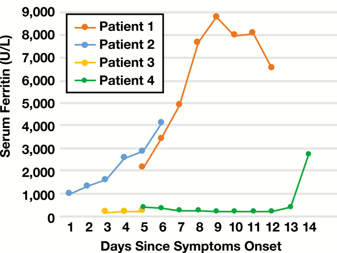
Objectives: A subset of coronavirus disease 2019 (COVID-19) patients exhibit clinical features of cytokine storm. However, clinicopathologic features diagnostic of hemophagocytic lymphohistiocytosis (HLH) have not been reported. We studied the reticuloendothelial organs of 4 consecutive patients who died of COVID-19 and correlated with clinical and laboratory parameters to detect HLH.

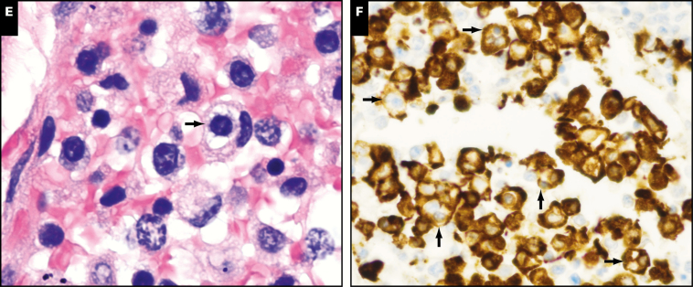
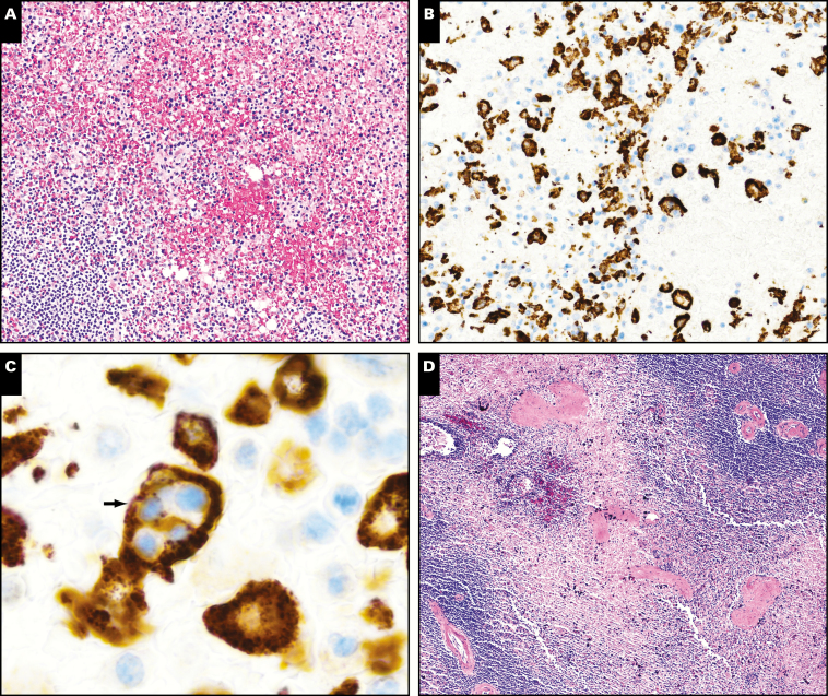
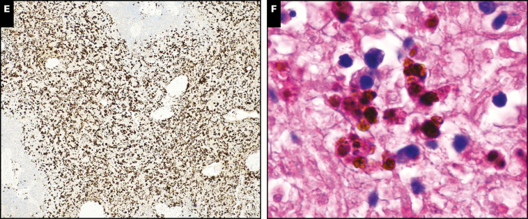
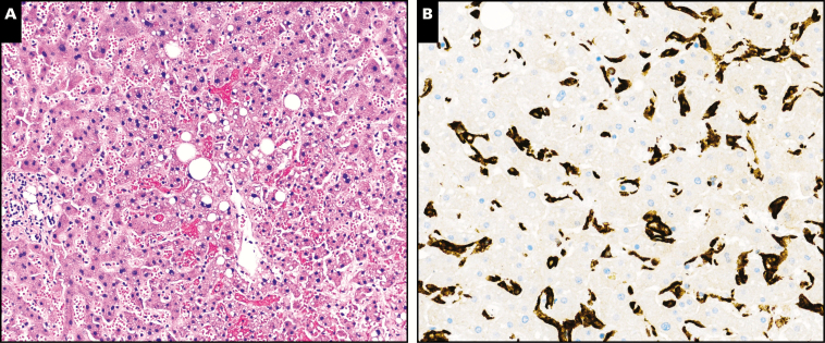
Methods: Autopsies were performed on 4 patients who died of COVID-19. Routine H&E staining and immunohistochemical staining for CD163 were performed to detect hemophagocytosis. Clinical and laboratory results from premortem blood samples were used to calculate H-scores.

Results: All 4 cases demonstrated diffuse alveolar damage within the lungs. Three of the 4 cases had histologic evidence of hemophagocytosis within pulmonary lymph nodes. One case showed hemophagocytosis in the spleen but none showed hemophagocytosis in liver or bone marrow. Lymphophagocytosis was the predominant form of hemophagocytosis observed. One patient showed diagnostic features of HLH with an H-score of 217, while a second patient likely had HLH with a partial H-score of 145 due to a missing triglyceride level. The remaining 2 patients had H-scores of 131 and 96.

Conclusions: This is the first report of severe acute respiratory syndrome coronavirus 2-associated HLH. Identification of HLH in a subset of patients with severe COVID-19 will inform clinical trials of therapeutic strategies.

Keywords: 2019-nCoV; Autopsy; COVID-19; Coronavirus; HLH; Hemophagocytic lymphohistiocytosis; Hemophagocytosis; SARS-CoV-2.


Figures

Image 1
Image 1
Hemophagocytosis in pulmonary hilar and mediastinal lymph nodes. H&E (A) and CD163 immunohistochemical (B) stains of lymph node from patient 1 showing distended cortical and subcortical sinusoids filled with histiocytes exhibiting focal necrosis (circled area). (×40). H&E (C) and CD163 immunohistochemical (D) stains of a lymph node from patient 2 showing a lesser degree of sinusoidal expansion, predominantly filling the subcapsular sinuses (circled areas) (×40). Hemophagocytosis consisted predominantly of lymphophagocytosis (arrow) in all cases and seen on H&E stain (E, ×1,000) and highlighted with CD163 immunohistochemical stain where numerous histiocytes phagocytosing 1 to several lymphocytes were apparent (arrows) (F, ×400).
Image 1
Image 1
Hemophagocytosis in pulmonary hilar and mediastinal lymph nodes. H&E (A) and CD163 immunohistochemical (B) stains of lymph node from patient 1 showing distended cortical and subcortical sinusoids filled with histiocytes exhibiting focal necrosis (circled area). (×40). H&E (C) and CD163 immunohistochemical (D) stains of a lymph node from patient 2 showing a lesser degree of sinusoidal expansion, predominantly filling the subcapsular sinuses (circled areas) (×40). Hemophagocytosis consisted predominantly of lymphophagocytosis (arrow) in all cases and seen on H&E stain (E, ×1,000) and highlighted with CD163 immunohistochemical stain where numerous histiocytes phagocytosing 1 to several lymphocytes were apparent (arrows) (F, ×400).
Image 2
Image 2
Hemophagocytosis in spleen. H&E stain of spleen from patient 2 with white pulp depletion and red pulp hemorrhage (A, ×100) with CD163 immunohistochemical stain demonstrating mild histiocytic hyperplasia (B, ×100) and lymphophagocytosis (arrow) (C, ×1,000). H&E stain of spleen from patient 1 showing mild white pulp depletion and red pulp infarction (D, ×40). CD163 immunohistochemical stain showing moderate histiocytic hyperplasia (E, ×40) and hemosiderin-laden macrophages suggestive of prior erythrophagocytosis (F, ×1,000).
Image 2
Image 2
Hemophagocytosis in spleen. H&E stain of spleen from patient 2 with white pulp depletion and red pulp hemorrhage (A, ×100) with CD163 immunohistochemical stain demonstrating mild histiocytic hyperplasia (B, ×100) and lymphophagocytosis (arrow) (C, ×1,000). H&E stain of spleen from patient 1 showing mild white pulp depletion and red pulp infarction (D, ×40). CD163 immunohistochemical stain showing moderate histiocytic hyperplasia (E, ×40) and hemosiderin-laden macrophages suggestive of prior erythrophagocytosis (F, ×1,000).
Image 3
Image 3
Liver without hemophagocytosis. A, H&E stain of liver showing mild centrilobular congestion with mild steatosis (×200). B, CD163 immunohistochemical stain showing mild Kupffer cell hyperplasia without hemophagocytosis (×200).
Figure 1
Figure 1
Serum ferritin levels over disease course.

References

    1. Johns Hopkins Coronavirus Resource Center. COVID-19 map https://coronavirus.jhu.edu/map.html. Accessed May 4, 2020.
    1. Arons MM, Hatfield KM, Reddy SC, et al. . Presymptomatic SARS-CoV-2 infections and transmission in a skilled nursing facility. N Engl J Med. 2020. doi:10.1056/NEJMoa2008457. - DOI - PMC - PubMed
    1. Pan Y, Yu X, Du X, et al. . Epidemiological and clinical characteristics of 26 asymptomatic SARS-CoV-2 carriers. J Infect Dis. 2020. doi:10.1093/infdis/jiaa205. - DOI - PMC - PubMed
    1. Li H, Liu L, Zhang D, et al. . SARS-CoV-2 and viral sepsis: observations and hypotheses. Lancet. 2020;395:1517-1520. doi:10.1016/S0140-6736(20)30920-X. - DOI - PMC - PubMed
    1. Mehta P, McAuley DF, Brown M, et al. . COVID-19: consider cytokine storm syndromes and immunosuppression. Lancet. 2020;395:1033-1034. doi:10.1016/S0140-6736(20)30628-0. - DOI - PMC - PubMed

Publication types

MeSH terms